Fraud Blocker Skip to main content

Human Bone Collection (page 6)

"Unveiling the Marvels of Human Bone: A Journey into our Remarkable Anatomy" Delving deep into the intricate wonders of human bone

Background imageHuman Bone Collection: Illustration of calcaneus, or heel bone, the three tarsal bones

Illustration of calcaneus, or heel bone, the three tarsal bones
Medicine: Human anatomy, calcaneus, or heel bone, the three tarsal bones. Drawing
Background imageHuman Bone Collection: Illustration of human muscles, biceps brachii muscle

Illustration of human muscles, biceps brachii muscle
Medicine: Human anatomy, muscles, biceps brachii muscle. Drawing

Background imageHuman Bone Collection: Illustration of human bones, patella, or knee cap, ballottement

Illustration of human bones, patella, or knee cap, ballottement
Medicine: Human anatomy, bones. Patella, or knee cap, ballottement. Drawing

Background imageHuman Bone Collection: Illustration of human pelvis, examples of damaged hips

Illustration of human pelvis, examples of damaged hips
Medicine: Human anatomy, pelvis, examples of damaged hips. Drawing

Background imageHuman Bone Collection: Illustration of apophysis in temporal and frontal bones

Illustration of apophysis in temporal and frontal bones
Medicine: Human anatomy, apophysis in the temporal and frontal bones. Drawing

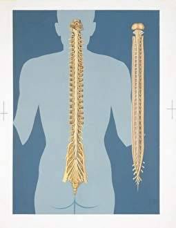
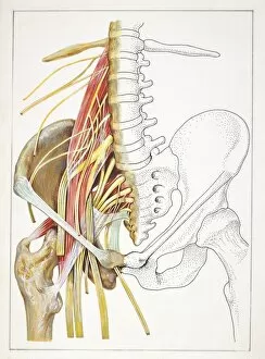
Background imageHuman Bone Collection: Illustration of human spinal cord

Illustration of human spinal cord
Medicine: Human anatomy, spinal cord. Drawing

Background imageHuman Bone Collection: Nervous system, spinal cord, drawing

Nervous system, spinal cord, drawing
Medicine - Anatomy - Nervous system - Spinal cord. Drawing

Background imageHuman Bone Collection: Illustration of human hip-joint

Illustration of human hip-joint
Medicine: Human anatomy, hip-joint. Drawing

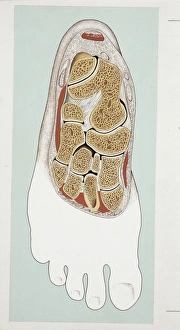
Background imageHuman Bone Collection: Illustration of human foot, horizontal section

Illustration of human foot, horizontal section
Medicine: Human anatomy, foot, horizontal section. Drawing

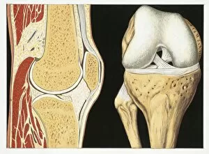
Background imageHuman Bone Collection: Illustration of human knee-joint

Illustration of human knee-joint
Medicine: Human anatomy, knee-joint. Drawing

Background imageHuman Bone Collection: Illustration of human bones, female type pelvis

Illustration of human bones, female type pelvis
Medicine: Human anatomy, bones. Female type pelvis. Drawing

Background imageHuman Bone Collection: Illustration of human bones, vertebrae articulations

Illustration of human bones, vertebrae articulations
Medicine: Human anatomy, bones. Vertebrae articulations. Drawing

Background imageHuman Bone Collection: Illustration of elbow articulation

Illustration of elbow articulation
Medicine: Human anatomy, elbow articulation. Drawing

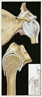
Background imageHuman Bone Collection: Illustration of left shoulder articulation

Illustration of left shoulder articulation
Medicine: Human anatomy, left shoulder articulation. Drawing

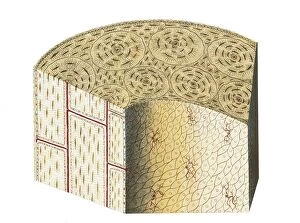
Background imageHuman Bone Collection: Illustration of human bone tissue

Illustration of human bone tissue
Medicine: Human anatomy, bone tissue, section. Drawing

Background imageHuman Bone Collection: Illustration of left shoulder blade

Illustration of left shoulder blade
Medicine: Human anatomy, left shoulder blade. Drawing

Background imageHuman Bone Collection: Illustration of muscles of the hand

Illustration of muscles of the hand
Medicine: Human anatomy, muscles of the hand. Drawing

Background imageHuman Bone Collection: Musculoskeletal (locomotor) system, muscles, drawing

Musculoskeletal (locomotor) system, muscles, drawing
Medicine - Anatomy - Musculoskeletal (locomotor) system - Muscles. Drawing

Background imageHuman Bone Collection: Illustration of lower limb

Illustration of lower limb
Medicine: Human anatomy, lower limb. Drawing

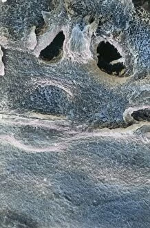
Background imageHuman Bone Collection: Bones, Osseous tissue under microscope

Bones, Osseous tissue under microscope
Medicine - Human Histology - Bones - Osseous tissue under microscope

Background imageHuman Bone Collection: Bones, Compact osseous tissue under microscope

Bones, Compact osseous tissue under microscope
Medicine - Human Histology - Bones - Compact osseous tissue under microscope

Background imageHuman Bone Collection: Bones, Connective tissue, Cartilage under microscope

Bones, Connective tissue, Cartilage under microscope
Medicine - Human Histology - Bones - Connective tissue - Cartilage under microscope

Background imageHuman Bone Collection: Bones, Connective tissue, Articular cartilage under microscope

Bones, Connective tissue, Articular cartilage under microscope
Medicine - Human Histology - Bones - Connective tissue - Articular cartilage under microscope

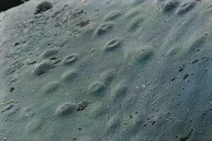
Background imageHuman Bone Collection: Bones, Osseous tissue of femur, extreme close-up

Bones, Osseous tissue of femur, extreme close-up
Medicine - Human Histology - Bones - Osseous tissue of a femur under microscope

Background imageHuman Bone Collection: Bones of the foot by Hieronymus Fabricius ab Aquapendente, anatomical plate

Bones of the foot by Hieronymus Fabricius ab Aquapendente, anatomical plate
Hieronymus Fabricius ab Aquapendente (1537-1619), Bones of the foot, anatomical plate

Background imageHuman Bone Collection: X-ray of arthritic knee

X-ray of arthritic knee
Medicine - Anatomy - Pathology - Diagnostic Imaging - X-ray of arthritic knee

Background imageHuman Bone Collection: Illustration of vertebral column and its possible movements

Illustration of vertebral column and its possible movements
Medicine: Human anatomy, vertebral column and its possible movements. Drawing

Background imageHuman Bone Collection: Male Neanderthal skull, front view

Male Neanderthal skull, front view

Background imageHuman Bone Collection: Babys skull with fontanelles showing, side view

Babys skull with fontanelles showing, side view

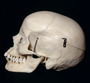
Background imageHuman Bone Collection: Female skull, side view

Female skull, side view

Background imageHuman Bone Collection: Musculoskeletal (locomotor) system, Vertebral column or spine, Osteoarthritis

Musculoskeletal (locomotor) system, Vertebral column or spine, Osteoarthritis, Deformation of the cervical spine
Medicine - Anatomy - Pathology - Diagnostic Imaging - Radiology - Orthopedics - Musculoskeletal (locomotor) system - Skeleton - Vertebral column or spine - Osteoarthritis - Deformation of

Background imageHuman Bone Collection: Illustration of lumbago, lumbar and pelvic regions

Illustration of lumbago, lumbar and pelvic regions
Medicine: lumbago, lumbar and pelvic regions. Drawing

Background imageHuman Bone Collection: View on musculoskeletal (locomotor) system, joints, bones, tibio-tarsal sprain, drawing

View on musculoskeletal (locomotor) system, joints, bones, tibio-tarsal sprain, drawing
Medicine - Anatomy - Pathology - Musculoskeletal (locomotor) system - Joints - Bones. Tibio-tarsal sprain. Drawing

Background imageHuman Bone Collection: Reconstruction of burial of Neanderthal Man (Homo sapiens neanderthalensis)

Reconstruction of burial of Neanderthal Man (Homo sapiens neanderthalensis) at La Chapelle-aux-Saints, France
Anthropology - Reconstruction of a burial of Neanderthal Man (Homo sapiens neanderthalensis) at La Chapelle-aux-Saints, France

Background imageHuman Bone Collection: Homo erectus pelvic bone fragments, from cave at Balzi Rossi cave complex, surroundings of Grimaldi

Homo erectus pelvic bone fragments, from cave at Balzi Rossi cave complex, surroundings of Grimaldi
Anthropology - Homo erectus pelvic bone fragments. From a cave at Balzi Rossi cave complex, surroundings of Grimaldi, province of Imperia

Background imageHuman Bone Collection: Illustration of whiplash injury

Illustration of whiplash injury

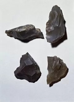
Background imageHuman Bone Collection: Bones from Archaeological area La Pineta, Isernia province, Molise region, Italy

Bones from Archaeological area La Pineta, Isernia province, Molise region, Italy
Prehistory, Paleolithic - Archaeological area La Pineta, Isernia province, Molise region, Italy

Background imageHuman Bone Collection: Skull of Neanderthal Man skeleton from burial in La Chapelle-aux-Saints

Skull of Neanderthal Man skeleton from burial in La Chapelle-aux-Saints
Prehistory, France, Middle Paleolithic. Burial of Neanderthal Man (Homo neanderthalensis) skeleton at La Chapelle-aux-Saints. Detail, skull

Background imageHuman Bone Collection: Skeleton known as Young Prince, from burial of Arene Candide Cave, in surroundings of Finale Ligure

Skeleton known as Young Prince, from burial of Arene Candide Cave, in surroundings of Finale Ligure
Prehistory, Italy, Upper Paleolithic. Skeleton known as the " Young Prince", from the burial of the Arene Candide Cave, in the surroundings of Finale Ligure, Savona province. Detail

Background imageHuman Bone Collection: Skeletons of woman and child from Bogebakken

Skeletons of woman and child from Bogebakken
Prehistory, Denmark, Mesolithic. Skeletons of a woman and a child. From Bogebakken

Background imageHuman Bone Collection: Herba Paris, illustration

Herba Paris, illustration
Manuscript, Italy, 15th century. Herbal from Trento. Plate: Herba Paris. Herb used to strengthen the bones. Manuscript 1591, folio 5, recto. Herbal with dedication to Saint Mark

Background imageHuman Bone Collection: Italy, Liguria Region, Caves of the Balzi Rossi. Cave of the Caviglione

Italy, Liguria Region, Caves of the Balzi Rossi. Cave of the Caviglione, Cro-Magnon type skull with shell headwear
Prehistory, Upper Paleolithic. Italy, Liguria Region - Caves of the Balzi Rossi. Cave of the Caviglione. Cro-Magnon type skeleton detail. Skull with headgear made of shells

Background imageHuman Bone Collection: Skeleton and funerary objects found in a burial site

Skeleton and funerary objects found in a burial site
Italy - Emilia Romagna Region - Villanovan period (900-720 BC). Skeleton and funerary objects found in a burial site

Background imageHuman Bone Collection: Fracture, computer artwork

Fracture, computer artwork
Bone fracture. Computer artwork showing detail of a bone fracture. The capillary (red) has remained intact. All fractures are reset into position and the bone heals naturally

Background imageHuman Bone Collection: Bone structure, macrophotograph

Bone structure, macrophotograph
Bone structure. Macrophotograph of a model of normal human bone structure

Background imageHuman Bone Collection: The bones of the feet, comuter artwork

The bones of the feet, comuter artwork
3d computer artwork of a side view of the bones of the feet

Background imageHuman Bone Collection: Spine and spinal nerves, computer artwork

Spine and spinal nerves, computer artwork
Lower part of the human vertebral column (blue) showing the spinal nerves (mauve).Through the spinal nerves messages from the brain to the body and from the body to the brain are relayed

Background imageHuman Bone Collection: The bones of the feet, computer artwork

The bones of the feet, computer artwork
3d computer artwork of a side view of the bones of the feet


All Professionally Made to Order for Quick Shipping

"Unveiling the Marvels of Human Bone: A Journey into our Remarkable Anatomy" Delving deep into the intricate wonders of human bone, we discover the backbone's unwavering strength, encompassing not only the resilient ribs but also the sturdy pelvis. Our skeletal framework, complemented by muscles that enable movement and support, forms a remarkable testament to nature's design. Transporting us back in time to 1896, an enchanting engraving reveals the distinct differences between male and female pelvises. This captivating depiction showcases how our bones adapt to accommodate childbirth and exemplifies their crucial role in sustaining life. Zooming closer to examine finer details, we explore the delicate yet robust structure of hand bones. These intricate marvels allow us to grasp objects with precision and dexterity—an essential characteristic that sets humans apart from other species. Shifting focus towards medical advancements, an anterior view displays a total shoulder joint repair—a testament to modern science's ability to mend even our most vital connections. Witnessing such interventions reminds us of both human resilience and our ongoing quest for better understanding and healing. Venturing further within ourselves, we encounter an exquisite engraving depicting organs' anatomy—a reminder that beneath these bones lie complex systems working harmoniously together—orchestrating life itself. The maxilla bone adorned with teeth serves as a gateway into oral health—the foundation upon which smiles are built. Its significance extends beyond aesthetics; it enables communication through speech while nourishing our bodies through mastication. A side view diagram unveils the intricacies of our spine—an architectural masterpiece supporting every movement we make. The cross-section diagram delves deeper into its connection with mouth and jaw structures—a reminder that each part is interconnected within this incredible vessel called "human. " Gazing at a mesmerizing engraving of a human skull transports us across centuries—reminding us of mortality while igniting curiosity about those who came before us.