
Human Bone Collection (page 2)
"Unveiling the Marvels of Human Bone: A Journey into our Remarkable Anatomy" Delving deep into the intricate wonders of human bone


All Professionally Made to Order for Quick Shipping
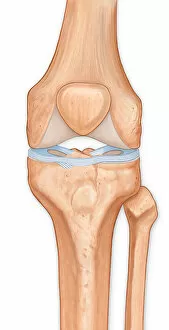
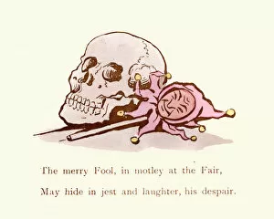
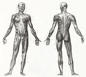
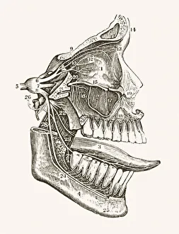
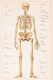
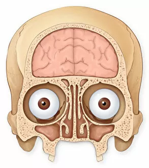
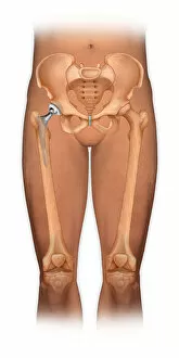
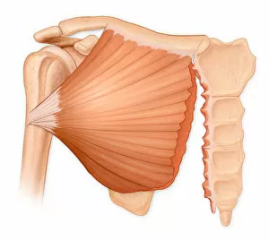
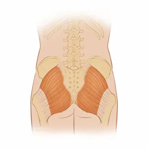
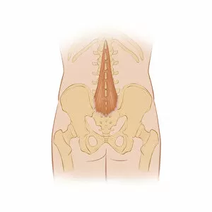
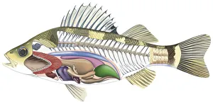
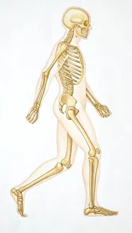
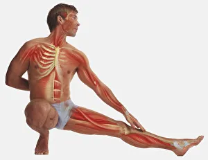
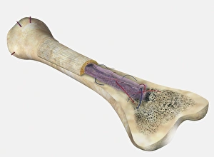
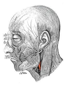
"Unveiling the Marvels of Human Bone: A Journey into our Remarkable Anatomy" Delving deep into the intricate wonders of human bone, we discover the backbone's unwavering strength, encompassing not only the resilient ribs but also the sturdy pelvis. Our skeletal framework, complemented by muscles that enable movement and support, forms a remarkable testament to nature's design. Transporting us back in time to 1896, an enchanting engraving reveals the distinct differences between male and female pelvises. This captivating depiction showcases how our bones adapt to accommodate childbirth and exemplifies their crucial role in sustaining life. Zooming closer to examine finer details, we explore the delicate yet robust structure of hand bones. These intricate marvels allow us to grasp objects with precision and dexterity—an essential characteristic that sets humans apart from other species. Shifting focus towards medical advancements, an anterior view displays a total shoulder joint repair—a testament to modern science's ability to mend even our most vital connections. Witnessing such interventions reminds us of both human resilience and our ongoing quest for better understanding and healing. Venturing further within ourselves, we encounter an exquisite engraving depicting organs' anatomy—a reminder that beneath these bones lie complex systems working harmoniously together—orchestrating life itself. The maxilla bone adorned with teeth serves as a gateway into oral health—the foundation upon which smiles are built. Its significance extends beyond aesthetics; it enables communication through speech while nourishing our bodies through mastication. A side view diagram unveils the intricacies of our spine—an architectural masterpiece supporting every movement we make. The cross-section diagram delves deeper into its connection with mouth and jaw structures—a reminder that each part is interconnected within this incredible vessel called "human. " Gazing at a mesmerizing engraving of a human skull transports us across centuries—reminding us of mortality while igniting curiosity about those who came before us.