
Homodimer Collection
A homodimer is a fascinating molecular structure that plays a crucial role in various biological processes


All Professionally Made to Order for Quick Shipping
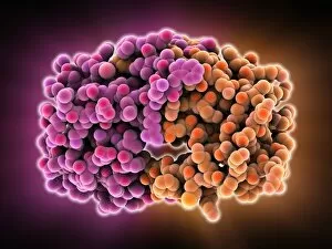
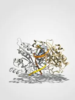
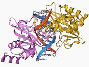
A homodimer is a fascinating molecular structure that plays a crucial role in various biological processes. One such example is the HIV-1 protease molecule, which forms a homodimer to carry out its vital function of cleaving viral polyproteins during replication. This dimeric arrangement allows for efficient and precise processing of these proteins, ultimately contributing to the maturation of infectious viral particles. Similarly, the EcoRV restriction enzyme molecule also exists as a homodimer. This enzyme acts as a defense mechanism in bacteria by recognizing specific DNA sequences and cutting them at precise locations. The dimeric form enhances its efficiency in target recognition and catalytic activity, ensuring accurate DNA cleavage. Within this family of EcoRV restriction enzymes, different variants like C014/2117, C014/2112, C014/2114, C014/2116, C014/2115, and F006/9496 have been identified. Each variant possesses distinct properties due to slight differences in their amino acid sequences or structural arrangements within the homodimeric complex. Moreover, scientists have extensively studied the interaction between HIV-1 protease molecules and inhibitors to develop effective antiviral drugs. By understanding how these inhibitors bind to each monomer within the homodimeric structure of HIV-1 protease molecules, researchers can design compounds that disrupt their enzymatic activity and hinder viral replication. In summary, whether it be through facilitating viral maturation or safeguarding bacterial genomes from foreign DNA invasion; homodimers like those formed by HIV-1 protease molecules or EcoRV restriction enzymes play critical roles in maintaining cellular integrity. Exploring their structures and functions not only deepens our knowledge of molecular biology but also paves the way for innovative therapeutic strategies against diseases caused by viruses or genetic abnormalities.