Fraud Blocker Skip to main content

Fluorescence Collection (page 3)

"Unveiling the Vibrant World of Fluorescence

Background imageFluorescence Collection: Adrenal gland, fluorescence micrograph C016 / 8472

Adrenal gland, fluorescence micrograph C016 / 8472
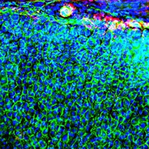
Adrenal gland. Fluorescence deconvolution micrograph of a section through an adrenal gland, showing the zona glomerulosa and zona fasciculata

Background imageFluorescence Collection: Kidney blood vessels, confocal micrograph C014 / 4610

Kidney blood vessels, confocal micrograph C014 / 4610
Kidney blood vessels. Laser scanning confocal micrograph (LSCM) of a section through a human kidney, showing a blood vessel (vertical, centre)

Background imageFluorescence Collection: Arabidopsis thaliana embryo, micrograph C014 / 4590

Arabidopsis thaliana embryo, micrograph C014 / 4590
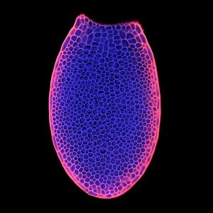
Arabidopsis thaliana embryo. Confocal light micrograph of a section through the embryo of a thale cress (Arabidopsis thaliana) plant

Background imageFluorescence Collection: Thale cress anther and pollen

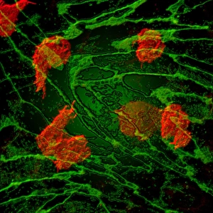
Thale cress anther and pollen. Confocal laser scanning micrograph of pollen grains (round) on a thale cress (Arabidopsis thaliana) anther

Background imageFluorescence Collection: Fibroblast cell, fluorescent micrograph

Fibroblast cell, fluorescent micrograph
Fibroblast cell. Confocal laser scanning micrograph of a fibroblast cell. The cell nucleus, which contains the cells genetic information, is green

Background imageFluorescence Collection: Plant protoplast, fluorescent micrograph

Plant protoplast, fluorescent micrograph
Protoplast. Confocal laser scanning micrograph of a tobacco plant (Nicotiana tabacum) protoplast. A protoplast is a plant cell that has had its tough outer cell wall removed by chemical treatment

Background imageFluorescence Collection: Plant trichome, fluorescent micrograph

Plant trichome, fluorescent micrograph
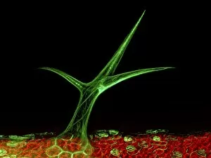
Plant trichome. Confocal laser scanning micrograph of a trichome (leaf hair) on a Arabidopsis thaliana plant. Actin filaments in the trichome are green

Background imageFluorescence Collection: Fluorite crystals C013 / 6697

Fluorite crystals C013 / 6697
Fluorite crystals. This sample is a mixture, and includes calcite, galena and pyrite. This specimen is from Glengowla East Mine, Oughterard, County Galway, Ireland

Background imageFluorescence Collection: Agate C013 / 6529

Agate C013 / 6529
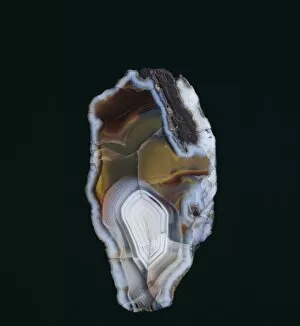
Agate. Section through a specimen of agate from Middlefield, Scotland, UK. Agate is an aggregate of different forms of silica (SiO2) within a geode

Background imageFluorescence Collection: HeLa cells, light micrograph C013 / 4774

HeLa cells, light micrograph C013 / 4774
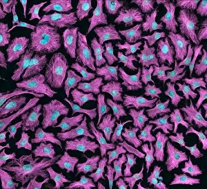
HeLa cells. Multi-photon fluorescence light micrograph of a group of cultured HeLa cells, showing the cell nuclei, which contain the cells genetic information (DNA, red)

Background imageFluorescence Collection: HeLa cells, light micrograph C013 / 4773

HeLa cells, light micrograph C013 / 4773
HeLa cells. Multi-photon fluorescence light micrograph of a group of cultured HeLa cells, showing the cell nuclei, which contain the cells genetic information (DNA, blue), and microtubules (pink)

Background imageFluorescence Collection: Natural ruby crystal

Natural ruby crystal
This ruby is a staggering 1, 085 carats. Mined in Burmas Mogok (Myanmar s) mines and bought by the Museum in 1924 from Burma Ruby Mines Ltd. It is one of the largest crystals in the Museum collection

Background imageFluorescence Collection: Susannite on Caledonite

Susannite on Caledonite
4mm crystal of susannite (carbonate) in a mass of caledonite (suplhate). Specimen from the Roughton Gill mine, Caldbeck Fells, Cumbria

Background imageFluorescence Collection: Fluorite, pale green octahedron

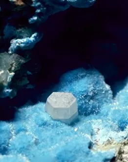
Fluorite, pale green octahedron
With calcite, galena and pyrite. Its properties include fluorescence and cubed crystals. Specimen from Glengowla East mine, Oughterard, Co. Galway, Eire

Background imageFluorescence Collection: Scheelite

Scheelite
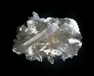
Crystals of scheelite (calcium tungstate), an important ore of tungsten from the collections of the Natural History Museum, London

Background imageFluorescence Collection: Diamonds fluorescing

Diamonds fluorescing
Murchison snuff box set with diamonds fluorescing under ultra violet radiation. Gold snuff box set with diamonds ranging from 0.75 to 2.5 carats, presented to Sir R. I. Murchison by Tsar Alexander II

Background imageFluorescence Collection: Celestite

Celestite
A mass of celestite crystals. Celestite is strontium sulphate and is the source of red colours in fireworks

Background imageFluorescence Collection: Autunite

Autunite comprises of (hydrated calcium uranyl phosphate). This is a green, radioactive, highly fluorescent mineral. This specimen is from the Natural History Museum, London

Background imageFluorescence Collection: Franklinite, fluorescing

Franklinite, fluorescing
Ore of zinc fluorescing under ultra violet radiation: calcite - pink; willemite - green; zincite - blue; franklinite - black. See image number 388 for white light view

Background imageFluorescence Collection: Willemite

Willemite
A specimen of willemite (zinc silicate) photographed under UV light

Background imageFluorescence Collection: Torbernite

Torbernite

Background imageFluorescence Collection: Opal doublets

Opal doublets
Opals are not truely crystalline and are therefore mineraloids. They comprise of (hydrated silica glass)

Background imageFluorescence Collection: Adamite

Adamite
A specimen of the mineral adamite (zinc arsenate hydroxide). It is a fluorescent mineral, popular among collectors and shows bright green fluorescent under UV lights

Background imageFluorescence Collection: Fluorescent group

Fluorescent group
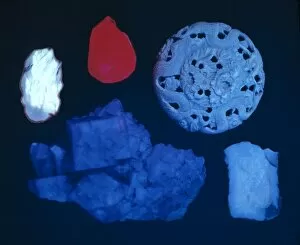
A group including amber, ruby, ivory, fluorite and opal photographed in ultra violet light. See 179 for the same group in white light

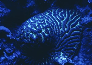
Background imageFluorescence Collection: Fluorescence emitted in corals, captured using special barrier filter. Tawali Resort

Fluorescence emitted in corals, captured using special barrier filter. Tawali Resort, Milne Bay, Papua New Guinea

Background imageFluorescence Collection: Natural occuring fluorescence in underwater soft coral (Sinularia sp. ), captured

Natural occuring fluorescence in underwater soft coral (Sinularia sp. ), captured
Natural occuring fluorescence in underwater soft coral (Sinularia sp.), captured by using special UV blocking barrier filters, Night Dive near Lewolin Village, Illi Api Island, Selat Boleng Strait

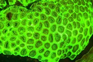
Background imageFluorescence Collection: Indonesia. Fluorsecense in coral (Goniopora sp. )

Indonesia. Fluorsecense in coral (Goniopora sp. )
Indonesia. Fluorsecense in coral (Goniopora sp.)

Background imageFluorescence Collection: Indonesia. Fluorescence in Tube Anemone (Ceranth idai)

Indonesia. Fluorescence in Tube Anemone (Ceranth idai)

Background imageFluorescence Collection: Natural occuring Fluorescence emitted and captured using special barrier filters

Natural occuring Fluorescence emitted and captured using special barrier filters over powerful underwater strobes. Indonesia, Raja Ampat region of Papua (formerly Irian Jaya)

Background imageFluorescence Collection: Fluorescing coral

Fluorescing coral

Background imageFluorescence Collection: X-ray viewing machine, 1895

X-ray viewing machine, 1895
X-ray viewing machine. Historical artwork of the fluoroscope designed and built by the French inventor Eugene Ducretet (1844-1915)

Background imageFluorescence Collection: Embryonic stem cells

Embryonic stem cells. Computer-enhanced confocal light micrograph of dividing stem cells from the ventricular zone of the retina of a developing embryo

Background imageFluorescence Collection: Dividing cell

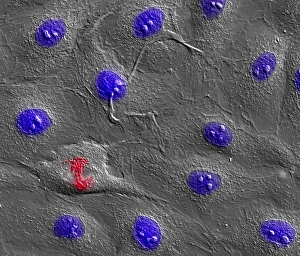
Dividing cell. Differential interference contrast (DIC) light micrograph of a cell (lower left) in the metaphase stage of mitosis (cell division). The cells nuclei are stained with fluorescent dye

Background imageFluorescence Collection: Dividing cell, light micrograph

Dividing cell, light micrograph
Cell division. Fluorescent light micrograph of a cell that has divided by mitosis, the asexual replication of a cell into two new cells

Background imageFluorescence Collection: Fibroblast cells, light micrograph

Fibroblast cells, light micrograph
Fibroblast cells. Confocal light micrograph of human fibroblast cells. The cell nuclei, which contain the cells genetic information, are red

Background imageFluorescence Collection: Brunners glands in the duodenum

Brunners glands in the duodenum
Brunners glands. Fluorescence confocal light micrograph of a section through a human duodenum (small intestine) showing Brunners glands (blue)

Background imageFluorescence Collection: Small intestine

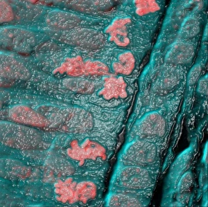
Small intestine. Fluorescence confocal light micrograph of a horizontal section through the mucosa of the human small intestine, showing crypts of Lieberkuhn (pink and blue)

Background imageFluorescence Collection: Colon lining

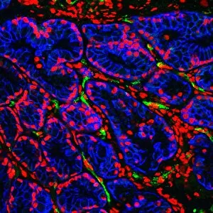
Colon lining. Fluorescence confocal light micrograph of the lining of a mouse colon (large intestine). The colon starts at the small intestine and ends at the rectum

Background imageFluorescence Collection: White blood cell response

White blood cell response
White blood cell movement. Computer-enhanced confocal light micrograph of white blood cells (red) moving through the intact walls of a blood vessel, a process known as diapedesis

Background imageFluorescence Collection: Crookes tube

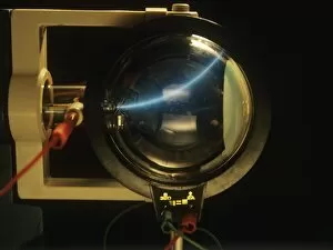
Crookes tube. Invented by William Crookes (1832 - 1919) in the late 19th century, this apparatus was used to investigate the path taken by cathode rays

Background imageFluorescence Collection: Bacterial contamination test

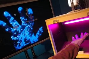
Bacterial contamination test. Ultraviolet (UV) light being used to reveal bacterial contamination. At right, a laboratory workers hand has been placed under a UV light source (pink)

Background imageFluorescence Collection: Cathode ray tube

Cathode ray tube. Image 2 of 2. Demonstration showing how a beam of electrons can be deflected by a magnetic field, as seen in television cathode ray tube (CRT) displays

Background imageFluorescence Collection: Banknote security

Banknote security. Euro banknote in ultraviolet light. This highlights security features intended to prevent fraud. Fluorescent ink and strips glow bright blue and red under ultraviolet light

Background imageFluorescence Collection: Blood sample testing

Blood sample testing. Medical technician preparing blood samples for a chemiluminescence immunity test. This test uses fluorescent antibodies to bind to antigens in the blood

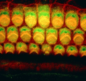
Background imageFluorescence Collection: Inner ear sensory hairs

Inner ear sensory hairs
Inner ear hair cells. Confocal light micrograph of V-shaped rows of hair cells (bright arcs) in the organ of Corti. This structure lies in the cochlea of the inner ear

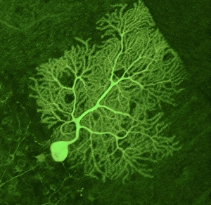
Background imageFluorescence Collection: Purkinje nerve cell, light micrograph

Purkinje nerve cell, light micrograph
Purkinje nerve cell. Confocal light micrograph of a purkinje cell (stained with green fluorescent protein), a type of neuron (nerve cell)

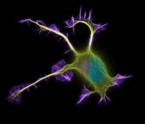
Background imageFluorescence Collection: Nerve cell growth

Nerve cell growth. Fluorescent light micrograph of a PC12 cell following stimulation by nerve growth factor. The cell body contains the nucleus (green)

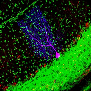
Background imageFluorescence Collection: Purkinje nerve cell

Purkinje nerve cell


All Professionally Made to Order for Quick Shipping

"Unveiling the Vibrant World of Fluorescence: Illuminating Cerebellum Tissue and Nerve Cells" Exploring the intricate beauty of cerebellum tissue through a mesmerizing light micrograph. Dive into the microscopic realm as nerve and glial cells come to life in a captivating light micrograph. Unraveling the secrets of glial cells with stunning clarity, thanks to a confocal light micrograph. Witness the enchanting glow of HeLa cells under a powerful microscope - an awe-inspiring sight captured in C017 / 8299. Delving deep into cell structure, fluorescence reveals hidden wonders that are otherwise invisible to our naked eyes. Marvel at the ethereal radiance emitted by HeLa cells, portrayed beautifully in C017 / 8298 - an image worth cherishing. Embark on a visual journey through cerebral cortex nerve cells, where fluorescence paints a vivid picture of their interconnectedness. Witness nature's miracle unfold as cell division is brought to life through an enchanting fluorescent micrograph. Behold another breathtaking view of cerebellum tissue illuminated by vibrant fluorescence - showcasing its complexity and elegance once again. Reflecting on history, discover how even X-ray security machines from 1900 were early pioneers in harnessing fluorescence for enhanced safety measures during screenings. Fluorescence unveils hidden worlds within ourselves and beyond – from delicate tissues to cellular processes – reminding us that there is always more than meets the eye.