Fraud Blocker Skip to main content

Fluorescence Collection (page 2)

"Unveiling the Vibrant World of Fluorescence

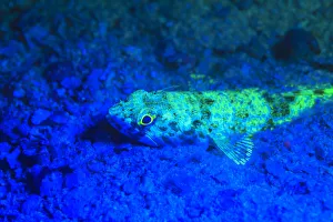
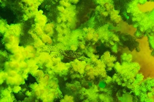
Background imageFluorescence Collection: Scorpionfish, Dusk Fluorescing, Red Sea, Egypt, Middle East

Scorpionfish, Dusk Fluorescing, Red Sea, Egypt, Middle East

Background imageFluorescence Collection: Day Fluorescing

Day Fluorescing

Background imageFluorescence Collection: Night dive at Barrier Reef near Saint Georges Caye, Fluorescence emitted at night

Night dive at Barrier Reef near Saint Georges Caye, Fluorescence emitted at night, exposed with special UV blocking filters, Belize, Central America

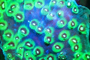
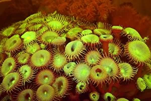
Background imageFluorescence Collection: Indo Pacific Sea Anemone, Day Fluorescing, Palau, Rock Islands-World Herititage Site

Indo Pacific Sea Anemone, Day Fluorescing, Palau, Rock Islands-World Herititage Site, Micronesia

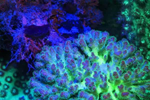
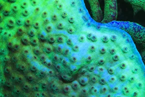
Background imageFluorescence Collection: Fused Staghorn Coral, Day Fluorescing, Palau, Rock Islands-World Herititage Site

Fused Staghorn Coral, Day Fluorescing, Palau, Rock Islands-World Herititage Site, Micronesia

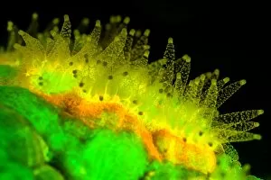
Background imageFluorescence Collection: Fluorescing Underwater Macro images

Fluorescing Underwater Macro images

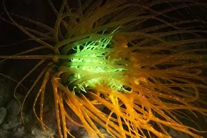
Background imageFluorescence Collection: Burrowing Tube Anemone, Night Fluorescing, Puerto Galera, Philippines

Burrowing Tube Anemone, Night Fluorescing, Puerto Galera, Philippines

Background imageFluorescence Collection: Picture No. 11676157

Picture No. 11676157
Soft Coral showing fluorescent colours when photographed under special blue light and filter photographed in aquarium Date:

Background imageFluorescence Collection: Electric discharges in rarefied gases. 2, 3, 4 and 6, Geissler tubes. 1 Fluorescence

Electric discharges in rarefied gases. 2, 3, 4 and 6, Geissler tubes. 1 Fluorescence of Sulphuret of calcium. 4 Nitrogen Vacuum (Spirals of Uranium Glass), 7, Hydrogen

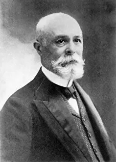
Background imageFluorescence Collection: (Antoine) Henri Becquerel (1852-1908) French physicist: Fluorescence: Radioactivity

(Antoine) Henri Becquerel (1852-1908) French physicist: Fluorescence: Radioactivity: shared 1903 Nobel prize for physics with Pierre and Marie Curie

Background imageFluorescence Collection: Jumping spider C018 / 4470

Jumping spider C018 / 4470
Jumping spider. Close-up photograph of a jumping spider specimen preserved in hand sanitiser solution. This specimen (species unknown) was found in the Dominican Republic

Background imageFluorescence Collection: Cotton stem, confocal micrograph C014 / 4636

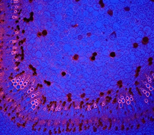
Cotton stem, confocal micrograph C014 / 4636
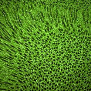
Cotton stem. Laser scanning confocal micrograph (LSCM) of a section through the stem of a cotton (Gossypium sp.) plant

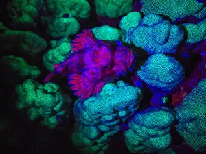
Background imageFluorescence Collection: Crab and coral fluorescing underwater C018 / 9373

Crab and coral fluorescing underwater C018 / 9373
Crab and coral fluorescing underwater under blue light. Some species of underwater animals emit a fluorescent glow. It is thought the glow may attract symbiotic algae

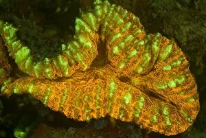
Background imageFluorescence Collection: Lobophyllia coral fluorescing C018 / 9375

Lobophyllia coral fluorescing C018 / 9375
Lobophyllia sp. coral fluorescing under blue light at night. Some species of coral emit a fluorescent glow. It is thought the glow may attract symbiotic algae

Background imageFluorescence Collection: Cerianthus anemone, fluorescing

Cerianthus anemone, fluorescing
Cerianthus anenome fluorescing under blue or ultraviolet light at night. Photographed in Borneo, Malaysia

Background imageFluorescence Collection: Reversibly switchable fluorescent protein

Reversibly switchable fluorescent protein, molecular model. Reversibly switchable fluorescent proteins (RSFPs) are proteins that can be repeatedly converted between a fluorescent

Background imageFluorescence Collection: Favites coral fluorescing

Favites coral fluorescing
Favites sp. coral polyps feeding at night, fluorescing bright orange, yellow and green when illuminated by blue light. Photographed in Borneo, Malaysia

Background imageFluorescence Collection: Squamous epithelium C014 / 4643

Squamous epithelium C014 / 4643
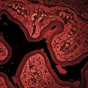
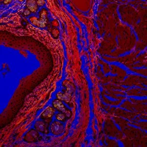
Squamous epithelium. Laser scanning confocal micrograph (LSCM) of a section through stratified squamous epithelial cells. Epithelial tissues line the cavities

Background imageFluorescence Collection: Squamous epithelium, confocal micrograph C014 / 4642

Squamous epithelium, confocal micrograph C014 / 4642
Squamous epithelium. Laser scanning confocal micrograph (LSCM) of a section through stratified squamous epithelial cells. Epithelial tissues line the cavities

Background imageFluorescence Collection: Lung tissue, confocal micrograph C014 / 4616

Lung tissue, confocal micrograph C014 / 4616
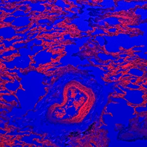
Lung tissue. Laser scanning confocal micrograph (LSCM) of a section through a human lung

Background imageFluorescence Collection: Kidney tissue, confocal micrograph C014 / 4611

Kidney tissue, confocal micrograph C014 / 4611
Kidney tissue. Laser scanning confocal micrograph (LSCM) of a section through a human kidney, showing the cell nuclei (dark)
Background imageFluorescence Collection: Liver portal triad, light micrograph C016 / 8490

Liver portal triad, light micrograph C016 / 8490
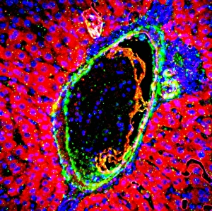
Liver portal triad. Fluorescence deconvolution micrograph of a section through a portal triad in liver tissue, showing hepatocyte cells (red)

Background imageFluorescence Collection: Liver portal triad, light micrograph C016 / 8489

Liver portal triad, light micrograph C016 / 8489
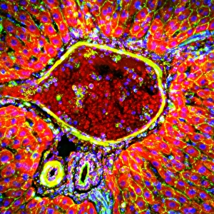
Liver portal triad. Fluorescence deconvolution micrograph of a section through a portal triad in liver tissue, showing hepatocyte cells (red)

Background imageFluorescence Collection: Liver portal triad, light micrograph C016 / 8488

Liver portal triad, light micrograph C016 / 8488
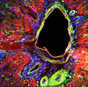
Liver portal triad. Fluorescence deconvolution micrograph of a section through a portal triad in liver tissue, showing hepatocyte cells (red)

Background imageFluorescence Collection: Kidney tissue, fluorescence micrograph C016 / 8484

Kidney tissue, fluorescence micrograph C016 / 8484
Kidney tissue. Fluorescence deconvolution micrograph of a section through a kidney, showing glomeruli (green), cell nuclei (blue dots), and renal tubules (red, circular)

Background imageFluorescence Collection: Heart muscle, fluorescence micrograph C016 / 8483

Heart muscle, fluorescence micrograph C016 / 8483
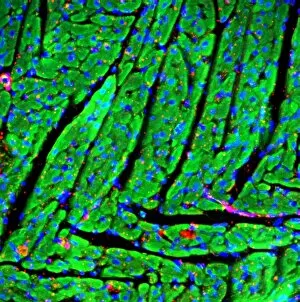
Heart muscle. Fluorescence deconvolution micrograph of a section through heart tissue, showing the angular distribution of the myocardium (cardiac muscle) fibres (green)

Background imageFluorescence Collection: Heart muscle, fluorescence micrograph C016 / 8481

Heart muscle, fluorescence micrograph C016 / 8481
Heart muscle. Fluorescence deconvolution micrograph of a section through heart tissue, showing blood vessels (oval, centre-left and upper centre) running through the myocardium (cardiac muscle)

Background imageFluorescence Collection: Heart muscle, fluorescence micrograph C016 / 8478

Heart muscle, fluorescence micrograph C016 / 8478
Heart muscle. Fluorescence deconvolution micrograph of a section through heart tissue, showing a blood vessel (diagonal, centre) running through the myocardium (cardiac muscle, green)

Background imageFluorescence Collection: Heart muscle, fluorescence micrograph C016 / 8477

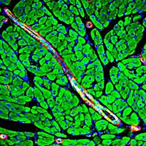
Heart muscle, fluorescence micrograph C016 / 8477
Heart muscle. Fluorescence deconvolution micrograph of a section through heart tissue, showing a blood vessel (blue, diagonal, centre) running through the myocardium (cardiac muscle, green)

Background imageFluorescence Collection: Adrenal gland, fluorescence micrograph C016 / 8472

Adrenal gland, fluorescence micrograph C016 / 8472
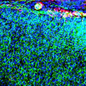
Adrenal gland. Fluorescence deconvolution micrograph of a section through an adrenal gland, showing the zona glomerulosa and zona fasciculata

Background imageFluorescence Collection: Kidney blood vessels, confocal micrograph C014 / 4610

Kidney blood vessels, confocal micrograph C014 / 4610
Kidney blood vessels. Laser scanning confocal micrograph (LSCM) of a section through a human kidney, showing a blood vessel (vertical, centre)

Background imageFluorescence Collection: Arabidopsis thaliana embryo, micrograph C014 / 4590

Arabidopsis thaliana embryo, micrograph C014 / 4590
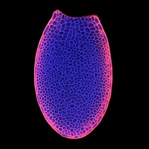
Arabidopsis thaliana embryo. Confocal light micrograph of a section through the embryo of a thale cress (Arabidopsis thaliana) plant

Background imageFluorescence Collection: Thale cress anther and pollen

Thale cress anther and pollen. Confocal laser scanning micrograph of pollen grains (round) on a thale cress (Arabidopsis thaliana) anther

Background imageFluorescence Collection: Fibroblast cell, fluorescent micrograph

Fibroblast cell, fluorescent micrograph
Fibroblast cell. Confocal laser scanning micrograph of a fibroblast cell. The cell nucleus, which contains the cells genetic information, is green

Background imageFluorescence Collection: Plant protoplast, fluorescent micrograph

Plant protoplast, fluorescent micrograph
Protoplast. Confocal laser scanning micrograph of a tobacco plant (Nicotiana tabacum) protoplast. A protoplast is a plant cell that has had its tough outer cell wall removed by chemical treatment

Background imageFluorescence Collection: Plant trichome, fluorescent micrograph

Plant trichome, fluorescent micrograph
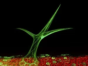
Plant trichome. Confocal laser scanning micrograph of a trichome (leaf hair) on a Arabidopsis thaliana plant. Actin filaments in the trichome are green

Background imageFluorescence Collection: Fluorite crystals C013 / 6697

Fluorite crystals C013 / 6697
Fluorite crystals. This sample is a mixture, and includes calcite, galena and pyrite. This specimen is from Glengowla East Mine, Oughterard, County Galway, Ireland

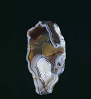
Background imageFluorescence Collection: Agate C013 / 6529

Agate C013 / 6529
Agate. Section through a specimen of agate from Middlefield, Scotland, UK. Agate is an aggregate of different forms of silica (SiO2) within a geode

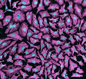
Background imageFluorescence Collection: HeLa cells, light micrograph C013 / 4774

HeLa cells, light micrograph C013 / 4774
HeLa cells. Multi-photon fluorescence light micrograph of a group of cultured HeLa cells, showing the cell nuclei, which contain the cells genetic information (DNA, red)

Background imageFluorescence Collection: HeLa cells, light micrograph C013 / 4773

HeLa cells, light micrograph C013 / 4773
HeLa cells. Multi-photon fluorescence light micrograph of a group of cultured HeLa cells, showing the cell nuclei, which contain the cells genetic information (DNA, blue), and microtubules (pink)

Background imageFluorescence Collection: Natural ruby crystal

Natural ruby crystal
This ruby is a staggering 1, 085 carats. Mined in Burmas Mogok (Myanmar s) mines and bought by the Museum in 1924 from Burma Ruby Mines Ltd. It is one of the largest crystals in the Museum collection

Background imageFluorescence Collection: Susannite on Caledonite

Susannite on Caledonite
4mm crystal of susannite (carbonate) in a mass of caledonite (suplhate). Specimen from the Roughton Gill mine, Caldbeck Fells, Cumbria

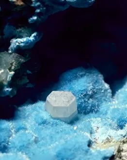
Background imageFluorescence Collection: Fluorite, pale green octahedron

Fluorite, pale green octahedron
With calcite, galena and pyrite. Its properties include fluorescence and cubed crystals. Specimen from Glengowla East mine, Oughterard, Co. Galway, Eire

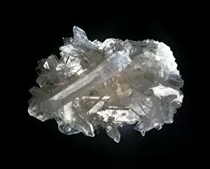
Background imageFluorescence Collection: Scheelite

Scheelite
Crystals of scheelite (calcium tungstate), an important ore of tungsten from the collections of the Natural History Museum, London

Background imageFluorescence Collection: Diamonds fluorescing

Diamonds fluorescing
Murchison snuff box set with diamonds fluorescing under ultra violet radiation. Gold snuff box set with diamonds ranging from 0.75 to 2.5 carats, presented to Sir R. I. Murchison by Tsar Alexander II

Background imageFluorescence Collection: Celestite

Celestite
A mass of celestite crystals. Celestite is strontium sulphate and is the source of red colours in fireworks

Background imageFluorescence Collection: Autunite

Autunite comprises of (hydrated calcium uranyl phosphate). This is a green, radioactive, highly fluorescent mineral. This specimen is from the Natural History Museum, London

Background imageFluorescence Collection: Franklinite, fluorescing

Franklinite, fluorescing
Ore of zinc fluorescing under ultra violet radiation: calcite - pink; willemite - green; zincite - blue; franklinite - black. See image number 388 for white light view


All Professionally Made to Order for Quick Shipping

"Unveiling the Vibrant World of Fluorescence: Illuminating Cerebellum Tissue and Nerve Cells" Exploring the intricate beauty of cerebellum tissue through a mesmerizing light micrograph. Dive into the microscopic realm as nerve and glial cells come to life in a captivating light micrograph. Unraveling the secrets of glial cells with stunning clarity, thanks to a confocal light micrograph. Witness the enchanting glow of HeLa cells under a powerful microscope - an awe-inspiring sight captured in C017 / 8299. Delving deep into cell structure, fluorescence reveals hidden wonders that are otherwise invisible to our naked eyes. Marvel at the ethereal radiance emitted by HeLa cells, portrayed beautifully in C017 / 8298 - an image worth cherishing. Embark on a visual journey through cerebral cortex nerve cells, where fluorescence paints a vivid picture of their interconnectedness. Witness nature's miracle unfold as cell division is brought to life through an enchanting fluorescent micrograph. Behold another breathtaking view of cerebellum tissue illuminated by vibrant fluorescence - showcasing its complexity and elegance once again. Reflecting on history, discover how even X-ray security machines from 1900 were early pioneers in harnessing fluorescence for enhanced safety measures during screenings. Fluorescence unveils hidden worlds within ourselves and beyond – from delicate tissues to cellular processes – reminding us that there is always more than meets the eye.