Fraud Blocker Skip to main content

Flowering Part Collection (page 3)

"Exploring the Intricate Beauty of Flowering Parts

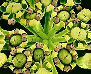
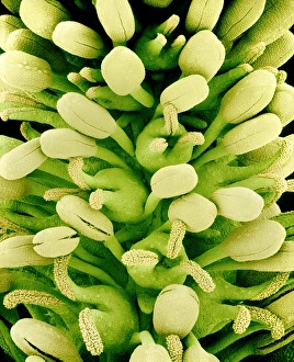
Background imageFlowering Part Collection: Fennel flowers, SEM

Fennel flowers, SEM
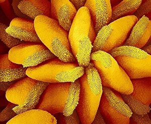
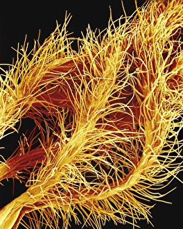
Fennel flower cluster. Coloured scanning electron micrograph (SEM) of a view from above of a cluster of fennel (Foeniculum vulgare) flowers

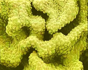
Background imageFlowering Part Collection: Melon pistil, SEM

Melon pistil, SEM
Melon flower pistil. Coloured scanning electron micrograph (SEM) of the surface of a melon flower (Cucumis melo) pistil. This is the female reproductive structure, composed of one or more carpels

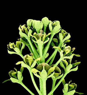
Background imageFlowering Part Collection: Field pennycress flowers, SEM

Field pennycress flowers, SEM
Field pennycress flowers. Coloured SEM (scanning electron micrograph) of the developing flowers of the field pennycress (Thlaspi arvense)

Background imageFlowering Part Collection: Sacred lotus anther, SEM

Sacred lotus anther, SEM
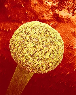
Sacred lotus anther. Coloured scanning electron micrograph (SEM) of the anther (male pollen- producing part) of a sacred lotus flower (Nelumbo nucifera). The anther opens when the pollen is mature

Background imageFlowering Part Collection: Carnation flower pistil, SEM

Carnation flower pistil, SEM
Carnation flower pistil. Coloured scanning electron micrograph (SEM) of the pistil of a carnation flower (Dianthus caryophyllus). The pistil is the female reproductive part of a flower

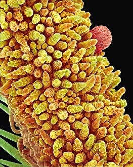
Background imageFlowering Part Collection: Morning glory stigma and pollen, SEM

Morning glory stigma and pollen, SEM
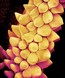
Morning glory stigma and pollen. Coloured scanning electron micrograph (SEM) of the stigma (brown) of a morning glory flower (Ipomoea sp.), with pollen grains (orange)

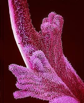
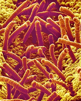
Background imageFlowering Part Collection: Periwinkle flower petal, SEM

Periwinkle flower petal, SEM
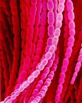
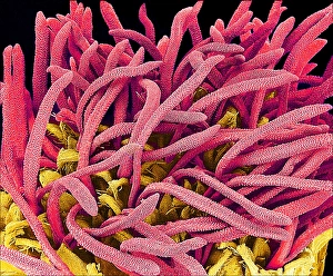
Periwinkle petal. Coloured scanning electron micrograph (SEM) of a periwinkle petal (family Apocynaceae). Its surface is covered in tiny epidermal hairs, or trichomes (pink)

Background imageFlowering Part Collection: Heath flower anthers, SEM

Heath flower anthers, SEM
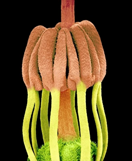
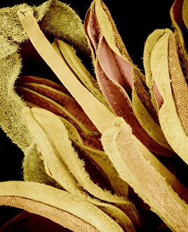
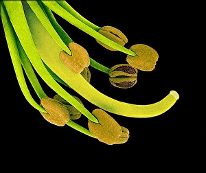
Heath flower stamens. Coloured scanning electron micrograph (SEM) of the stamens of a flower of a heath plant (Erica sp.)

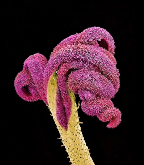
Background imageFlowering Part Collection: Primrose stigma, SEM

Primrose stigma, SEM
Primrose stigma. Coloured scanning electron micrograph (SEM) of the stigma (yellow) and style (lower left) of a primrose flower (Primula sp.)

Background imageFlowering Part Collection: Buttercup flower pistils, SEM

Buttercup flower pistils, SEM
Buttercup flower pistil. Coloured scanning electron micrograph (SEM) of buttercup (Ranunculus sp.) flower pistils. A pistil is the female reproductive structure, composed of one or more carpels

Background imageFlowering Part Collection: Oleander flower anthers, SEM

Oleander flower anthers, SEM
Oleander flower anthers. Coloured scanning electron micrograph (SEM) of the five anthers of the flower of an oleander plant (Nerium sp.)

Background imageFlowering Part Collection: Flowers, SEM

Flowers, SEM
Reproductive flower parts. Coloured scanning electron micrograph (SEM) of unidentified flowers. The sepals (green) surround and protect central reproductive parts (yellow)

Background imageFlowering Part Collection: Cranesbill pistil, SEM

Cranesbill pistil, SEM
Cranesbill pistil. Coloured scanning electron micrograph (SEM) of the surface of the pistil of a cranesbill plant (Geranium sp.). The pistil is the female reproductive part of a flower

Background imageFlowering Part Collection: Flower stigma, SEM

Flower stigma, SEM
Flower stigma. Coloured scanning electron micrograph (SEM) of a flowers stigma (yellow) and style (green). These are part of a plants female reproductive structures

Background imageFlowering Part Collection: Flower reproductive parts, SEM

Flower reproductive parts, SEM
Flower reproductive parts. Coloured SEM (scanning electron micrograph) of the reproductive parts of an unidentified flower

Background imageFlowering Part Collection: Comfrey petals, SEM

Comfrey petals, SEM
Comfrey petals. Coloured scanning electron micrograph (SEM) of the petals of a comfrey (Symphytum officinale) flower. Comfrey is a perennial herb with large

Background imageFlowering Part Collection: Pansy petal, SEM

Pansy petal, SEM
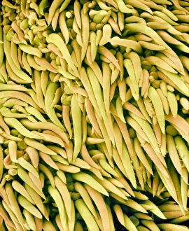
Pansy petal. Coloured scanning electron micrograph (SEM) of the surface of a pansy flower petal (Viola tricolor). The petal is covered in tiny epidermal hairs (cone-shaped objects)

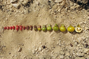
Background imageFlowering Part Collection: Buds and fruits of the wild pomegranate

Buds and fruits of the wild pomegranate
Wild pomegranate reproduction. Buds (far left), flowers (centre left), and developing fruits (centre to far right) of the wild pomegranate tree (Punica protopunica)

Background imageFlowering Part Collection: Withered flowers, SEM

Withered flowers, SEM
Withered flowers. Coloured scanning electron micrograph (SEM) of dying flowers of the softstem bulrush (Schoenoplectus tabernaemontani) plant. Magnification: x12 at 6x7cm size

Background imageFlowering Part Collection: Flower bud of the snapdragon

Flower bud of the snapdragon
False-colour scanning electron micrograph of a flower bud of the snapdragon, Antirrhinum majus. The sepals of the flower have been removed to show the compact structure of the bud early in its

Background imageFlowering Part Collection: Pumpkin flower petal, SEM

Pumpkin flower petal, SEM
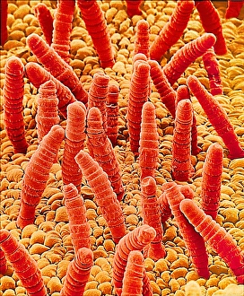
Pumpkin petal. Coloured scanning electron micrograph (SEM) of the surface of a pumpkin petal (Cucurbita sp. ). The petal is covered in tiny epidermal hairs (trichomes, brown)

Background imageFlowering Part Collection: Geranium flower stigma, SEM

Geranium flower stigma, SEM
Geranium flower stigma. Coloured scanning electron micrograph (SEM) of the stigma of a geranium flower (Geranium thunbergii)

Background imageFlowering Part Collection: Bellflower pistil, SEM

Bellflower pistil, SEM
Bellflower pistil. Coloured scanning electron micrograph (SEM) of the pistil of a bellflower plant (Campanula sp.). The pistil is the female reproductive part of a flower

Background imageFlowering Part Collection: Stamens and carpels, SEM

Stamens and carpels, SEM
Stamens and carpels. Coloured scanning electron micrograph (SEM) of the stamens (male, yellow) and carpels (female) of the Houttuynia cordata plant

Background imageFlowering Part Collection: Plant pores, SEM

Plant pores, SEM
Plant pores. Coloured scanning electron micrograph (SEM) of stomata (pores) on the pistil of a bellflower (Adenophora sp.). Stomata are the site of gaseous exchange, or respiration

Background imageFlowering Part Collection: Tulip anther, light micrograph

Tulip anther, light micrograph
Tulip anther. Light micrograph of a cross-section through the anther of a tulip bud (Tulipa sp.), showing a full anther sac with developing pollen grains (white)

Background imageFlowering Part Collection: Pumpkin petal, SEM

Pumpkin petal, SEM
Pumpkin petal. Coloured scanning electron micrograph (SEM) of the surface of a pumpkin petal (Cucurbita sp. ). The petal is covered in tiny epidermal hairs (trichomes)

Background imageFlowering Part Collection: Female butternut squash flower

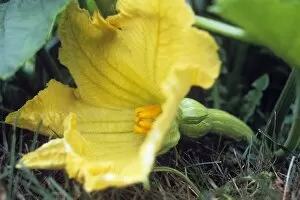
Female butternut squash flower (Cucurbita moschata). Plants in the Cucurbitaceae family are monoecious, meaning they have unisexual (imperfect) flowers of both sexes on the same plant

Background imageFlowering Part Collection: Whiteweed flower pistils, SEM

Whiteweed flower pistils, SEM
Whiteweed flower floret pistils. Coloured scanning electron micrograph (SEM) of the pistils (red, the female reproductive organs) emerging from the true flowers or florets (yellow)

Background imageFlowering Part Collection: Tulips reproductive structures

Tulips reproductive structures. The female part of the flower is the carpel, at the top of which is the stigma. This is supported on the style

Background imageFlowering Part Collection: Pea flower stigma, SEM

Pea flower stigma, SEM
Pea flower stigma. Coloured scanning electron micrograph (SEM) of the surface of a common or garden pea flower (Pisum sativum) stigma

Background imageFlowering Part Collection: Pollen tube, light micrograph

Pollen tube, light micrograph
Pollen tube. Light micrograph of a section through an evening primrose (Oenthera biennis) stigma. A pollen grain (circular)

Background imageFlowering Part Collection: Judas tree flower, SEM

Judas tree flower, SEM
Judas tree flower. Coloured scanning electron micrograph (SEM) of the reproductive parts of the flower of the Judas tree, or redbud (Cercis sp.)

Background imageFlowering Part Collection: Daisy flower petals, SEM

Daisy flower petals, SEM
Daisy flower petals. Coloured scanning electron micrograph (SEM) of drooping petals from an ox- eye daisy (Chrysanthemum leucanthemum) flower head


All Professionally Made to Order for Quick Shipping

"Exploring the Intricate Beauty of Flowering Parts: A Captivating SEM Journey" Delve into the mesmerizing world of flowering parts as we embark on a captivating scanning electron microscope (SEM) adventure. Witness the intricate details and stunning structures that make up these botanical wonders. The Easter cactus stigma, magnified through SEM, reveals its delicate and feathery texture, showcasing nature's artistry at its finest. The Tea flower stamens come alive under the microscope, displaying their vibrant colors and fascinating patterns that attract pollinators with precision. A closer look at the Buttercup flower exposes its velvety petals in exquisite detail. Each petal seems to dance with light, inviting us to appreciate its ephemeral beauty. Meanwhile, the humble Chickweed flower surprises us with an unexpected burst of color when viewed through false-color SEM imaging techniques. Discovering hidden secrets within flowers continues as we observe oil glands on peppermint petals glistening like tiny droplets of gold. The Aubergine flower petal captivates our senses with its unique shape and intricate surface patterns revealed by SEM technology. Lily petals unfold before our eyes in breathtaking clarity under SEM examination; their graceful curves and delicate veins become works of art themselves. Columbine flower stamens reveal their complex arrangement, designed to ensure successful reproduction for this remarkable species. Diving deeper into nature's marvels, a false-color SEM image unveils the enchanting intricacies of a chickweed flower – each component playing a vital role in attracting pollinators while ensuring survival for generations to come. Yarrow's petal surface becomes an abstract masterpiece when observed through an electron microscope lens - every ridge and groove telling tales of resilience against harsh environments. And finally, witness the ovary of Opium poppy in astonishing detail; this microscopic view offers insights into how life begins within these magnificent blooms.