
Embryology Collection
Embryology, the study of the development of embryos from fertilization to birth, has been a fascinating field for centuries


All Professionally Made to Order for Quick Shipping
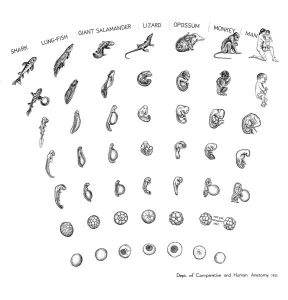
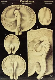
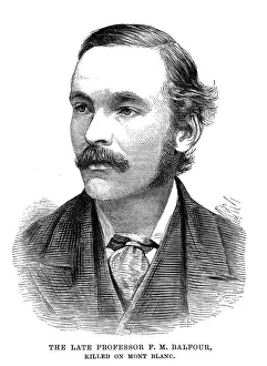
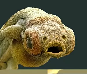
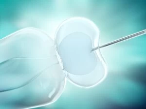
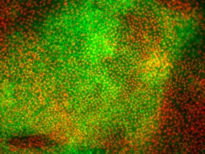
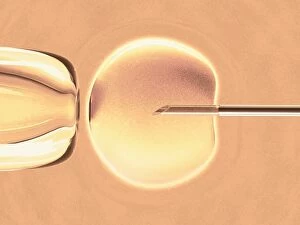
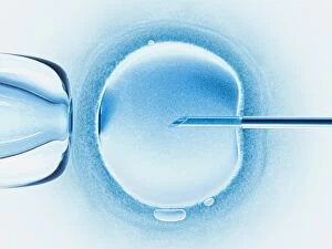
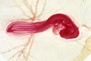
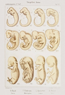
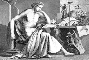
Embryology, the study of the development of embryos from fertilization to birth, has been a fascinating field for centuries. From ancient philosophers like Aristotle to modern-day scientists, it has captivated our curiosity and deepened our understanding of life itself. In 1932, the Department of Comparative and Human Anatomy at the American Museum of Natural History created a groundbreaking chart showcasing comparative embryology. This chart traced the developmental journey from a fish embryo all the way to a human being, highlighting striking similarities and subtle differences along the way. The beauty of this scientific discipline lies in its ability to reveal connections between different species. Artwork depicting fish embryos alongside salamanders, tortoises, and chicks further emphasizes these shared patterns among diverse organisms. Throughout history, notable figures have contributed significantly to embryological research. Geoffroy Saint-Hilaire's engraving captures his dedication to unraveling nature's secrets. Karl Ernst von Baer, a renowned German biologist from the 19th century, made significant contributions that still resonate today. Girolamo Fabrici stands as an influential figure in Italian anatomical studies during the 17th century. His work not only laid foundations for embryology but also advanced microscopical anatomy and histology. Mammal embryos depicted in 1905 serve as visual evidence supporting evolutionary theories proposed by scientists like Professor Francis Maitland Balfour. His Scottish origins did not hinder him from leaving an indelible mark on embryological research before his untimely death in 1882. Another luminary in this field was Karl Ernst von Baer—a German naturalist who hailed originally from Estonia—whose meticulous observations led him to formulate fundamental principles known as "Baer's laws. " Marcello Malpighi is revered as one of history's most prominent biologists and physicians; he earned recognition as the "Father of microscopical anatomy, histology physiology, and embryology.