Fraud Blocker Skip to main content

Embryo Collection (page 8)

Embryo - a tiny marvel of life, the beginning of a new existence

Background imageEmbryo Collection: Eight-cell embryo, artwork F006 / 2170

Eight-cell embryo, artwork F006 / 2170
Morula embryo. Computer artwork of a 16-cell morula embryo

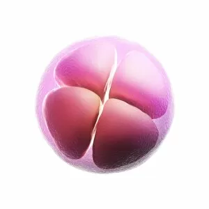
Background imageEmbryo Collection: Four-cell embryo, artwork F006 / 2169

Four-cell embryo, artwork F006 / 2169
Four-cell embryo, computer artwork

Background imageEmbryo Collection: Two-cell embryo, artwork F006 / 2167

Two-cell embryo, artwork F006 / 2167
Two-cell embryo, computer artwork

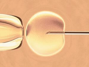
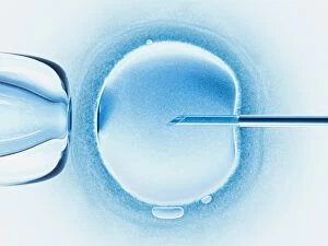
Background imageEmbryo Collection: In vitro fertilisation, artwork F006 / 8779

In vitro fertilisation, artwork F006 / 8779
In vitro fertilisation, computer artwork

Background imageEmbryo Collection: In vitro fertilisation, artwork F006 / 8778

In vitro fertilisation, artwork F006 / 8778
In vitro fertilisation, computer artwork

Background imageEmbryo Collection: Embryo, artwork F006 / 9906

Embryo, artwork F006 / 9906
Embryo, computer artwork

Background imageEmbryo Collection: Embryo, artwork F006 / 9904

Embryo, artwork F006 / 9904
Embryo, computer artwork

Background imageEmbryo Collection: Embryo, artwork F006 / 9905

Embryo, artwork F006 / 9905
Embryo, computer artwork

Background imageEmbryo Collection: Chicken embryo, light micrograph F006 / 9811

Chicken embryo, light micrograph F006 / 9811
Chicken embryo. Light micrograph of a chicken (Gallus domesticus) embryo 36 hours after fertilisation

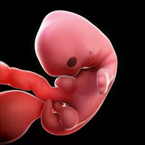
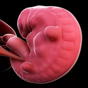
Background imageEmbryo Collection: Embryo at 7 weeks, artwork F006 / 7887

Embryo at 7 weeks, artwork F006 / 7887
Embryo at 7 weeks, computer artwork

Background imageEmbryo Collection: Embryo at 6 weeks, artwork F006 / 7886

Embryo at 6 weeks, artwork F006 / 7886
Embryo at 6 weeks, computer artwork

Background imageEmbryo Collection: Embryo at 5 weeks, artwork F006 / 7885

Embryo at 5 weeks, artwork F006 / 7885
Embryo at 5 weeks, computer artwork

Background imageEmbryo Collection: Embryo at 4 weeks, artwork F006 / 7884

Embryo at 4 weeks, artwork F006 / 7884
Embryo at 4 weeks, computer artwork

Background imageEmbryo Collection: Dodder seed, SEM C015 / 5077

Dodder seed, SEM C015 / 5077
Dodder (Cuscuta sp.) sede, coloured environmental scanning electron micrograph (ESEM)

Background imageEmbryo Collection: Developing fish fin, SEM

Developing fish fin, SEM
Developing fish fin. Coloured scanning electron micrograph (SEM) of a fin from a zebrafish (Danio rerio) embryo. Zebrafish are freshwater fish that are found in India

Background imageEmbryo Collection: Mouse embryo, prepared specimen C018 / 0318

Mouse embryo, prepared specimen C018 / 0318
Mouse embryo. Macrophotograph of a prepared specimen of a mouse embryo after around 16 days of development. It has been made translucent and its skeleton stained with Alizarin red dye

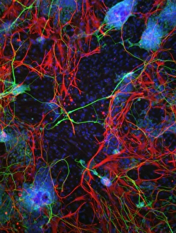
Background imageEmbryo Collection: Stem cell-derived nerve cells

Stem cell-derived nerve cells. Fluorescence light micrograph of neural (nerve) stem cells that have been derived from human embryonic stem cells (HESC)

Background imageEmbryo Collection: Ectopic pregnancy, artwork C016 / 8737

Ectopic pregnancy, artwork C016 / 8737
Ectopic pregnancy, artwork. This is a pregnancy where the embryo (upper left) implants outside the uterus, usually as here in the Fallopian tube that should channel the fertilised egg to the uterus

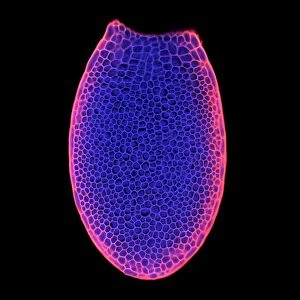
Background imageEmbryo Collection: Arabidopsis thaliana embryo, micrograph C014 / 4590

Arabidopsis thaliana embryo, micrograph C014 / 4590
Arabidopsis thaliana embryo. Confocal light micrograph of a section through the embryo of a thale cress (Arabidopsis thaliana) plant

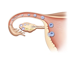
Background imageEmbryo Collection: Ovulation and fertilisation, artwork

Ovulation and fertilisation, artwork
Ovulation and fertilisation. Computer artwork of the female reproductive system, showing the development of an ovum (egg, small circle) inside an ovary (oval)

Background imageEmbryo Collection: Nucleoplasmin histone-chaperone protein C015 / 6915

Nucleoplasmin histone-chaperone protein C015 / 6915
Nucleoplasmin histone-chaperone protein, molecular model. This is nucleoplasmin 2 (Npm2), a chaperone that acts on human nucleoplasmin. Chaparone proteins play a key role in aiding protein folding

Background imageEmbryo Collection: Redwood seeds, SEM C015 / 5169

Redwood seeds, SEM C015 / 5169
Redwood (Sequoia sp.) seeds, coloured environmental scanning electron micrograph (ESEM)

Background imageEmbryo Collection: Redwood seeds, SEM C015 / 5170

Redwood seeds, SEM C015 / 5170
Redwood (Sequoia sp.) seeds, coloured environmental scanning electron micrograph (ESEM)

Background imageEmbryo Collection: Corn germination, SEM C015 / 5166

Corn germination, SEM C015 / 5166
Corn germination. Coloured environmental scanning electron micrograph (ESEM) of a germinating corn (Zea mays) seedling. The radicle (green), the primary root has emerged

Background imageEmbryo Collection: Zebra fish embryo, SEM C013 / 9586

Zebra fish embryo, SEM C013 / 9586
Zebra fish embryo. Coloured scanning electron micrograph (SEM) of the embryo of a zebrafish (Danio rerio), showing its eyes either side of its large open mouth (centre)

Background imageEmbryo Collection: Chickens egg outer shell detail, SEM

Chickens egg outer shell detail, SEM
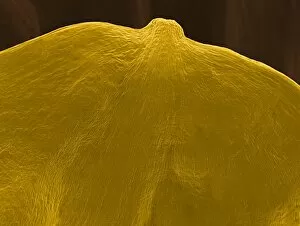
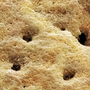
Chickens egg outer shell detail, coloured scanning electron micrograph (SEM). Hard-shelled eggs are produced by birds and some reptiles. The shell protects the growing embryo inside

Background imageEmbryo Collection: Germinated seed, SEM

Germinated seed, SEM
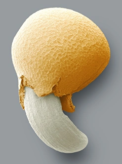
Germinated seed. Image 4 of 4. Coloured scanning electron micrograph (SEM) of the final stage in the germination of a plant seed. The seed coat (testa) has been lost

Background imageEmbryo Collection: Germinating seed, SEM

Germinating seed, SEM
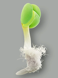
Germinating seed. Image 1 of 4. Coloured scanning electron micrograph (SEM) of one of the earliest stages of the germination of a plant seed

Background imageEmbryo Collection: Aptenodytes fosteri, emperor penguin egg

Aptenodytes fosteri, emperor penguin egg
Collected on the British Terra Nova Antarctic Expedition (1910 - 1913) by Edward Wilson

Background imageEmbryo Collection: Lizard embryo

Lizard embryo
Model of a lizard embryo

Background imageEmbryo Collection: Aptenodytes forsteri, Emperor penguin eggs

Aptenodytes forsteri, Emperor penguin eggs
Eggs of Aptenodytes forsteri (Emperor penguin) brought back from Apsley Cherry-Garrards 1911 Antarctic expedition. The holes were made to allow investigation of the embryos

Background imageEmbryo Collection: Protoceratops embryo

Protoceratops embryo

Background imageEmbryo Collection: Drodromeus Makelei Embryo DInosaur - artist conception based on cat scan

Drodromeus Makelei Embryo DInosaur - artist conception based on cat scan
FG-7258 Drodromeus Makelei Embryo DInosaur - artist conception based on cat scan Francois Gohier contact details: prints@ardea.com tel: +44 (0) 20 8318 1401

Background imageEmbryo Collection: Domestic Sheep, Texel tup lambs, all produced by same sire, born as embryo transplants

Domestic Sheep, Texel tup lambs, all produced by same sire, born as embryo transplants, standing in pasture, England, may

Background imageEmbryo Collection: Domestic Sheep, recipient mule ewe with pedigree Beltex lamb, produced as embryo transplant

Domestic Sheep, recipient mule ewe with pedigree Beltex lamb, produced as embryo transplant, standing in pasture, England, may


All Professionally Made to Order for Quick Shipping

Embryo - a tiny marvel of life, the beginning of a new existence. In the realm of human biology, the term "embryo" refers to a stage in early development known as the human blastocyst. This remarkable phase marks the formation of various cell types that will eventually give rise to all organs and tissues. But humans are not alone in this journey from conception to birth. Comparative embryology sheds light on our shared ancestry with other species. A fascinating chart created by the Department of Comparative and Human Anatomy at the American Museum of Natural History in 1932 showcases this connection. From fish to man, it reveals striking similarities in embryonic development across different organisms. Take, for instance, a chicken chick nestled within its eggshell after twenty days of incubation. At this point, feathers begin to emerge as delicate wings take shape beneath their protective shell. Similarly captivating is an image capturing developing trout eggs; each one holds immense potential for growth and survival. Pictures numbered 11675528 through 11675525 offer glimpses into these diverse embryos' intricate structures and stages of maturation—a testament to nature's incredible diversity and complexity. Meanwhile, another snapshot displays a ten-day-old chicken embryo still enclosed within its egg—an awe-inspiring sight showcasing how life unfolds even before hatching occurs. Zooming closer into human reproduction, we encounter an astonishing photograph revealing embryo development just 24-36 hours after fertilization (Picture No. 12019792). It captures those crucial initial moments when cells rapidly divide and multiply—laying down foundations for future growth. Lastly, let us not forget about our own kind—the miracle that takes place inside every woman's womb: a human fetus cradled within its amniotic sac (Picture No. XXXXXXXX). As it floats weightlessly amidst nourishing fluids, vital organs form gradually while tiny limbs grow stronger day by day—a testament to both resilience and vulnerability.