Fraud Blocker Skip to main content

Embryo Collection (page 5)

Embryo - a tiny marvel of life, the beginning of a new existence

Background imageEmbryo Collection: Picture No. 10875787

Picture No. 10875787
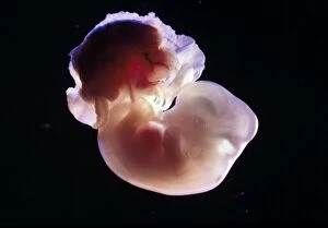
Light Micrograph of Rat Embryo - 14.5 days into its gestation period - in sac with placenta Date:

Background imageEmbryo Collection: Picture No. 10875793

Picture No. 10875793
Light Micrograph of Rat Embryo without its yolk sac, at 17.5 days Date:

Background imageEmbryo Collection: Picture No. 10875789

Picture No. 10875789
Light Micrograph of Rat Embryo at 14.5days old Date:

Background imageEmbryo Collection: Picture No. 10875791

Picture No. 10875791
Light Micrograph of Rat Embryo at 15.5 days, no sac, no placenta Date:

Background imageEmbryo Collection: Picture No. 10875788

Picture No. 10875788
Light Micrograph of Rat Embryo - 14.5 days into its gestation period - in sac with placenta Date:

Background imageEmbryo Collection: Picture No. 10875785

Picture No. 10875785
Light Micrograph of Rat Embryo 13.2 days after fertilisation Date:

Background imageEmbryo Collection: Picture No. 10875782

Picture No. 10875782
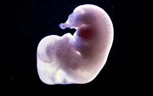
Light Micrograph of Rat Embryo 11.5 days after fertilisation Date:

Background imageEmbryo Collection: Picture No. 10875786

Picture No. 10875786
Light Micrograph of Rat Embryo 13.2 days after fertilisation, out of its yolk sac Date:

Background imageEmbryo Collection: Picture No. 10875780

Picture No. 10875780
Light Micrograph of Rat Embryo 11.5 days after fertilisation Date:

Background imageEmbryo Collection: Picture No. 10875783

Picture No. 10875783
Light Micrograph of Rat Embryo 13.2 days after fertilisation Date:

Background imageEmbryo Collection: Picture No. 10875784

Picture No. 10875784
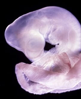
Light Micrograph of Eye detail of a rat embryo at 13.2 days after fertilisation Date:

Background imageEmbryo Collection: Picture No. 10875781

Picture No. 10875781
Light Micrograph of Rat Embryo 11.5 days after fertilisation Date:

Background imageEmbryo Collection: Picture No. 10875779

Picture No. 10875779
Light Micrograph of Rat Embryo 10.2 days after fertilisation in sac Date:

Background imageEmbryo Collection: Picture No. 10875778

Picture No. 10875778
Light Micrograph of Human Embryo With Placenta Date:

Background imageEmbryo Collection: Picture No. 10865272

Picture No. 10865272
Birth of Tammar wallaby (Macropus eugenii) embryo enters pouch after a 2 minute climb. Australia Date:

Background imageEmbryo Collection: Spitzer Space Telescope false-colour image of mountains where stars are born

Spitzer Space Telescope false-colour image of mountains where stars are born. Dubbed Mountains of Creation the towering pillars of cool gas and dust are lit at the tips with light from embryo stars

Background imageEmbryo Collection: MR. DISRAELIs VISIT TO GLASGOW: 1. The Crowd outside the Kibble Palace. 2. Embryo

MR. DISRAELIs VISIT TO GLASGOW: 1. The Crowd outside the Kibble Palace. 2. Embryo Divines amusing themselves. 3. Cheering the Lord Rector. 4. Capping the Lord Rector. 5. Mr. Disraeli at the Desk. 6

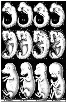
Background imageEmbryo Collection: Comparison of embryos of Pig, Cow, Rabbit and Man

Comparison of embryos of Pig, Cow, Rabbit and Man
Haeckels comparison of embryos of Pig, Cow, Rabbit and Man. Top row, all embryos show gill slit at O, demonstrating his Recapitulation theory

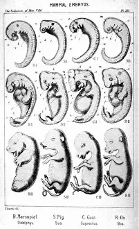
Background imageEmbryo Collection: Mammal embryos

Mammal embryos. From Ernst Haeckel The Evolution of Man, fifth edition, London, 1910

Background imageEmbryo Collection: Sauropsida embryos

Sauropsida embryos. From Ernst Haeckel The Evolution of Man, fifth edition, London, 1910

Background imageEmbryo Collection: Cattle farming, inserting embryo transfer gun into recipient cow, England, April

Cattle farming, inserting embryo transfer gun into recipient cow, England, April

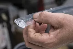
Background imageEmbryo Collection: Cattle farming, bottle of anaesthetic for epidural into recipient cows during embryo transfer

Cattle farming, bottle of anaesthetic for epidural into recipient cows during embryo transfer, England, April

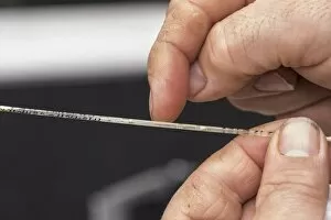
Background imageEmbryo Collection: Cattle farming, straw containing embryo for embryo transfer to recipient cows, England, April

Cattle farming, straw containing embryo for embryo transfer to recipient cows, England, April

Background imageEmbryo Collection: Cattle farming, removing straws of embryos from flask containing liquid nitrogen for embryo

Cattle farming, removing straws of embryos from flask containing liquid nitrogen for embryo transfer into recipient cows, England, April

Background imageEmbryo Collection: Cattle farming, end of embryo transfer gun, England, April

Cattle farming, end of embryo transfer gun, England, April

Background imageEmbryo Collection: Cattle farming, filling syringe from bottle of anaesthetic for epidural into recipient cows during

Cattle farming, filling syringe from bottle of anaesthetic for epidural into recipient cows during embryo transfer, England, April

Background imageEmbryo Collection: Developmental biology, Embryology, Human embryo in amniotic sac at ninth week of gestation

Developmental biology, Embryology, Human embryo in amniotic sac at ninth week of gestation
Medicine - Anatomy - Developmental biology - Embryology - Human embryo in the amniotic sac at ninth week of gestation

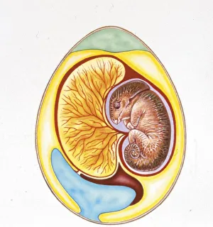
Background imageEmbryo Collection: Illustration of cross section of dinosaur egg

Illustration of cross section of dinosaur egg
Palaeozoology - Section of a dinosaur egg - Art work

Background imageEmbryo Collection: Above view of 3 Eggs

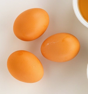
Above view of 3 Eggs

Background imageEmbryo Collection: Model of chicken embryo developing inside egg

Model of chicken embryo developing inside egg

Background imageEmbryo Collection: Model of day-old birds egg with network of blood vessels developing

Model of day-old birds egg with network of blood vessels developing

Background imageEmbryo Collection: Model of freshly-laid birds egg

Model of freshly-laid birds egg

Background imageEmbryo Collection: Model of 3-day-old birds egg with embryo beginning to grow

Model of 3-day-old birds egg with embryo beginning to grow

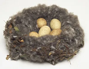
Background imageEmbryo Collection: Overhead view of the nest, containing five eggs, of a Red-Breasted Goose

Overhead view of the nest, containing five eggs, of a Red-Breasted Goose

Background imageEmbryo Collection: Ostrich Egg

Ostrich Egg

Background imageEmbryo Collection: Slug inside egg

Slug inside egg

Background imageEmbryo Collection: Bird embryo attached to yolk sac

Bird embryo attached to yolk sac

Background imageEmbryo Collection: Great crested newt (Triturus cristatus), embryo developing inside egg

Great crested newt (Triturus cristatus), embryo developing inside egg

Background imageEmbryo Collection: A tiny bird embryo in a glass dish

A tiny bird embryo in a glass dish

Background imageEmbryo Collection: Developing embryo of Great Crested Newt (Triturus cristatus) on leaf

Developing embryo of Great Crested Newt (Triturus cristatus) on leaf

Background imageEmbryo Collection: Anterior-posterior polarity, illustration C018 / 0916

Anterior-posterior polarity, illustration C018 / 0916
Anterior-posterior polarity, illustration. The unequal distribution of proteins or other factors within a newly fertilised zygote or developing embryo is a key mechanism for establishing the axes

Background imageEmbryo Collection: In vitro fertilisation, artwork F006 / 3726

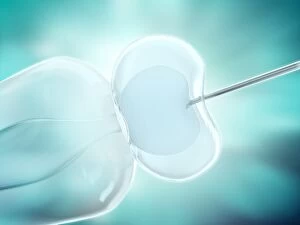
In vitro fertilisation, artwork F006 / 3726
In vitro fertilisation, computer artwork

Background imageEmbryo Collection: Blastocyst embryo, light micrograph F008 / 3573

Blastocyst embryo, light micrograph F008 / 3573
Blastocyst embryo, light micrograph. This embryo is five to six days old


All Professionally Made to Order for Quick Shipping

Embryo - a tiny marvel of life, the beginning of a new existence. In the realm of human biology, the term "embryo" refers to a stage in early development known as the human blastocyst. This remarkable phase marks the formation of various cell types that will eventually give rise to all organs and tissues. But humans are not alone in this journey from conception to birth. Comparative embryology sheds light on our shared ancestry with other species. A fascinating chart created by the Department of Comparative and Human Anatomy at the American Museum of Natural History in 1932 showcases this connection. From fish to man, it reveals striking similarities in embryonic development across different organisms. Take, for instance, a chicken chick nestled within its eggshell after twenty days of incubation. At this point, feathers begin to emerge as delicate wings take shape beneath their protective shell. Similarly captivating is an image capturing developing trout eggs; each one holds immense potential for growth and survival. Pictures numbered 11675528 through 11675525 offer glimpses into these diverse embryos' intricate structures and stages of maturation—a testament to nature's incredible diversity and complexity. Meanwhile, another snapshot displays a ten-day-old chicken embryo still enclosed within its egg—an awe-inspiring sight showcasing how life unfolds even before hatching occurs. Zooming closer into human reproduction, we encounter an astonishing photograph revealing embryo development just 24-36 hours after fertilization (Picture No. 12019792). It captures those crucial initial moments when cells rapidly divide and multiply—laying down foundations for future growth. Lastly, let us not forget about our own kind—the miracle that takes place inside every woman's womb: a human fetus cradled within its amniotic sac (Picture No. XXXXXXXX). As it floats weightlessly amidst nourishing fluids, vital organs form gradually while tiny limbs grow stronger day by day—a testament to both resilience and vulnerability.