
Differential Interference Contrast Collection
"Differential Interference Contrast: Revealing the Hidden Beauty of Microscopic Worlds" Step into the mesmerizing realm (DIC) microscopy


All Professionally Made to Order for Quick Shipping
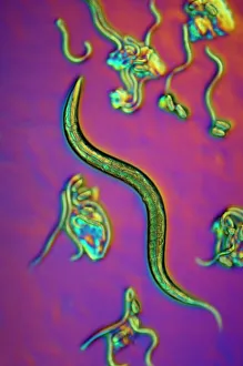
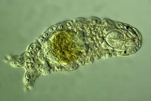
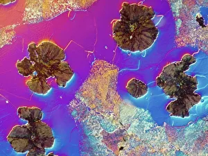
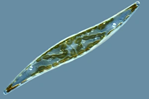
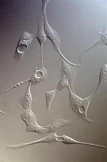
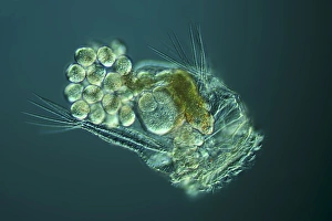
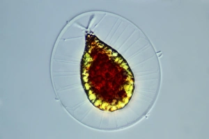
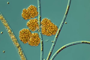
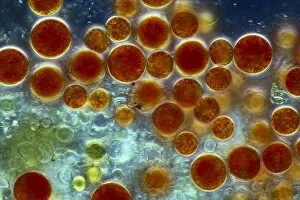
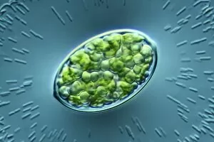
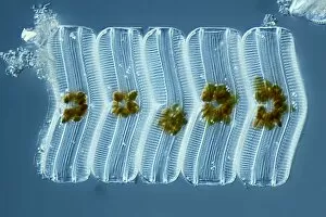
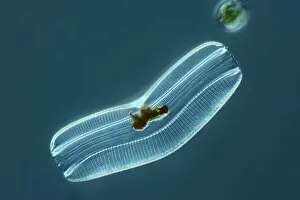
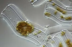
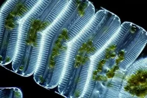
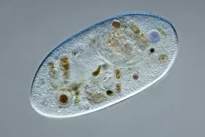
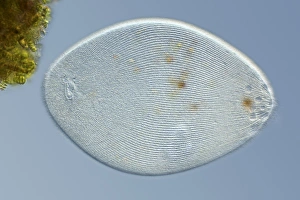
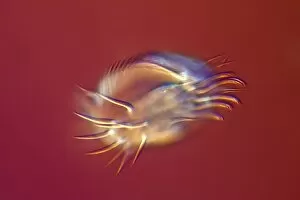
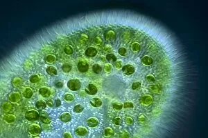
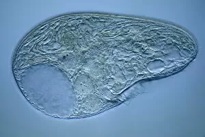
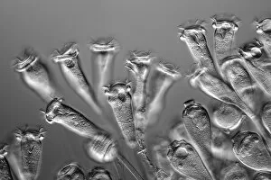
"Differential Interference Contrast: Revealing the Hidden Beauty of Microscopic Worlds" Step into the mesmerizing realm (DIC) microscopy, where a whole new world unfolds before our eyes. This advanced imaging technique allows us to explore intricate details and structures that are otherwise invisible to the naked eye. In one captivating light micrograph, we witness the enchanting dance of C. Elegans worms. Their delicate bodies shimmer with vibrant colors, showcasing their remarkable beauty and complexity. Moving on, we encounter a water bear captured in another light micrograph. Its translucent body reveals an astonishing level of intricacy as every tiny feature is brought to life through DIC. The sheer resilience and adaptability of this extraordinary creature become evident in this stunning image. A fascinating glimpse into the microscopic world continues with a light micrograph capturing cast iron under DIC illumination. The intricate patterns and textures within this seemingly mundane material come alive, transforming it into a work of art. Diatoms take center stage in yet another breathtaking light micrograph. These single-celled organisms exhibit unparalleled symmetry and grace as they float effortlessly through their aquatic habitat. MDCK cells appear inverted but no less captivating in their DIC portrayal. Every cell membrane becomes distinct, allowing us to appreciate the complex network that forms these living structures. The wonders continue with a rotifer delicately cradling its eggs under DIC illumination. This tender moment showcases both fragility and strength within nature's cycle of life. A midge larva emerges from obscurity thanks to DIC microscopy, revealing its unique anatomy and features previously hidden from view. Transitioning from light microscopy to scanning electron microscopy (SEM), we delve into the realm of amoeboid protozoa – creatures whose shape-shifting abilities astound us all over again at such magnification levels. Returning back to light micrographs, Haematococcus alga takes center stage once more; its vivid red hue contrasting against a backdrop of green.