Fraud Blocker Skip to main content

Diagnostic Collection (page 6)

"Unlocking the Secrets of Health: Exploring the World Tools" Medieval Urine Wheel: From ancient times to modern medicine

Background imageDiagnostic Collection: Calcified heart valve, CT scan

Calcified heart valve, CT scan
Calcified heart valve. Coloured computed tomography (CT) scan of a section through the heart of a 67-year-old patient showing calcification of the aortic valve (round)

Background imageDiagnostic Collection: Kidney tumour, CT scan

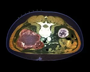
Kidney tumour, CT scan
Kidney tumour. Coloured computed tomography (CT) scan of a section through the abdomen of a 62-year-old patient, showing necrosis (death) of tissue in the right kidney

Background imageDiagnostic Collection: Calcified heart valve, CT scans

Calcified heart valve, CT scans
Calcified heart valve. Coloured computed tomography (CT) scans of a section through the heart of a 67-year-old patient showing calcification of the aortic valve (round)

Background imageDiagnostic Collection: Normal skull, 3D CT scan C016 / 6330

Normal skull, 3D CT scan C016 / 6330
Normal skull. Coloured cut-away 3D computed tomography (CT) scan of the healthy skull of a 35 year old patient

Background imageDiagnostic Collection: Human head, 3D CT scan C016 / 6339

Human head, 3D CT scan C016 / 6339
Human head. Coloured 3D computed tomography (CT) scan of a healthy human head. The soft tissues are shown at right, the skull is seen at left

Background imageDiagnostic Collection: Normal head and neck, 3D CT scan C016 / 6335

Normal head and neck, 3D CT scan C016 / 6335
Normal head. Coloured 3D computed tomography (CT) scan of the healthy head and neck of a 35 year old patient

Background imageDiagnostic Collection: Normal head and neck, 3D CT scan C016 / 6334

Normal head and neck, 3D CT scan C016 / 6334
Normal head. Coloured 3D computed tomography (CT) scan of the healthy head and neck of a 35 year old patient

Background imageDiagnostic Collection: Normal head and neck, MRI and 3D CT scans C016 / 6338

Normal head and neck, MRI and 3D CT scans C016 / 6338
Normal head. Coloured composite image of a magnetic resonance imaging (MRI) scan of the brain and a 3D computed tomography (CT) scan of the head and neck of a 35 year old patient

Background imageDiagnostic Collection: Normal head and neck, MRI and 3D CT scans C016 / 6336

Normal head and neck, MRI and 3D CT scans C016 / 6336
Normal head. Coloured composite image of a magnetic resonance imaging (MRI) scan of the brain and a 3D computed tomography (CT) scan of the head and neck of a 35 year old patient

Background imageDiagnostic Collection: Normal skull, 3D CT scan C016 / 6328

Normal skull, 3D CT scan C016 / 6328
Normal skull. Coloured 3D computed tomography (CT) scan of the healthy skull of a 35 year old patient

Background imageDiagnostic Collection: Normal skull, 3D CT scan C016 / 6326

Normal skull, 3D CT scan C016 / 6326
Normal skull. Coloured 3D computed tomography (CT) scan of the healthy skull of a 35 year old patient

Background imageDiagnostic Collection: Normal skull, 3D CT scan C016 / 6331

Normal skull, 3D CT scan C016 / 6331
Normal skull. Coloured cut-away 3D computed tomography (CT) scan of the healthy skull of a 35 year old patient

Background imageDiagnostic Collection: Human head, 3D CT scan C016 / 6393

Human head, 3D CT scan C016 / 6393
Human head. Coloured 3D computed tomography (CT) scan of a profile view of a 30 year old patients head and neck

Background imageDiagnostic Collection: Normal skull, 3D CT scan C016 / 6327

Normal skull, 3D CT scan C016 / 6327
Normal skull. Coloured 3D computed tomography (CT) scan of the healthy skull of a 35 year old patient

Background imageDiagnostic Collection: Human head, 3D CT scan C016 / 6377

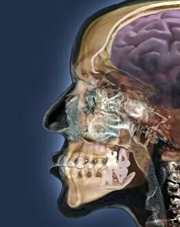
Human head, 3D CT scan C016 / 6377
Human head. Coloured 3D computed tomography (CT) scan of a profile view of a 47 year old patients head. The brain is purple and bone is light brown. At centre, behind the face, are the sinuses

Background imageDiagnostic Collection: Lung silicosis, light micrograph C015 / 7129

Lung silicosis, light micrograph C015 / 7129
Lung silicosis. Light micrograph of a section through a lung with silicosis, a type of pneumoconiosis caused by exposure to silica dusts. The lung tissue has become fibrous (centre)

Background imageDiagnostic Collection: Stethoscope demonstration C015 / 8835

Stethoscope demonstration C015 / 8835
MODEL RELEASED. Stethoscope demonstration. Medical worker showing how a stethoscope can be used to listen to your own heartbeat

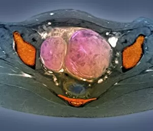
Background imageDiagnostic Collection: Uterine fibroids, MRI scan C018 / 0467

Uterine fibroids, MRI scan C018 / 0467
Uterine fibroids. Coloured magnetic resonance imaging (MRI) scan of the pelvis of a 47 year old female with two large fibroids (pink) in her uterus

Background imageDiagnostic Collection: Polycystic kidneys, CT scans C018 / 0571

Polycystic kidneys, CT scans C018 / 0571
Polycystic kidneys. Coloured computed tomography (CT) scans of the abdomen of a 28 year old patient with polycystic kidneys (pink)

Background imageDiagnostic Collection: Rectal cancer, MRI scans C018 / 0508

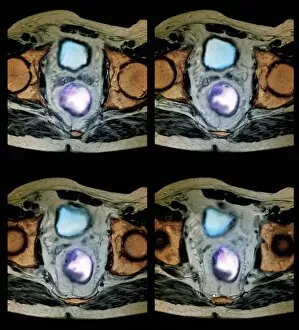
Rectal cancer, MRI scans C018 / 0508
Rectal cancer. Coloured magnetic resonance imaging (MRI) scans of the pelvis of a 65 year old patient with a malignant (cancerous) tumour (purple) in the rectum, the last part of the large intestine

Background imageDiagnostic Collection: Rectal cancer, MRI scans C018 / 0510

Rectal cancer, MRI scans C018 / 0510
Rectal cancer. Coloured magnetic resonance imaging (MRI) scans of the pelvis of a 65 year old patient with a malignant (cancerous) tumour in the rectum, the last part of the large intestine

Background imageDiagnostic Collection: Polycystic kidneys, CT scans C018 / 0572

Polycystic kidneys, CT scans C018 / 0572
Polycystic kidneys. Coloured computed tomography (CT) scans of the abdomen of a 28 year old patient with polycystic kidneys (dark blue)

Background imageDiagnostic Collection: Trachea, 3D CT scan C018 / 0394

Trachea, 3D CT scan C018 / 0394
Trachea. Coloured 3D computed tomography (CT) scan of the healthy trachea (windpipe) of a 45 year old patient at the point that it bifurcates into the two primary bronchi (airways)

Background imageDiagnostic Collection: Spinal cord cyst, MRI scan C018 / 0538

Spinal cord cyst, MRI scan C018 / 0538
Spinal cord cyst. Magnetic resonance imaging (MRI) scan of the cervical (neck) spinal cord of a 28 year old patient with syringomyelia, a cyst in the spinal cord. A cyst is a fluid-filled sac

Background imageDiagnostic Collection: Heart and lung disease, X-rays C018 / 0502

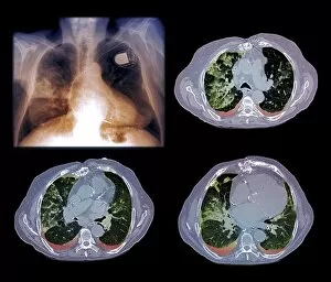
Heart and lung disease, X-rays C018 / 0502
Heart and lung disease. Coloured X-ray (top left) and computed tomography (CT) scans of the chest of an 80 year old patient with heart and lung disease

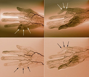
Background imageDiagnostic Collection: Blood supply of the hand, angiograms C018 / 0389

Blood supply of the hand, angiograms C018 / 0389
Blood supply of the hand. Coloured angiograms (X-rays) of the blood vessels of the right hand of a 65 year old patient with Dupuytrens contracture

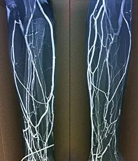
Background imageDiagnostic Collection: Forearm veins, X-ray C018 / 0475

Forearm veins, X-ray C018 / 0475
Forearm veins. Coloured venogram (vein X-ray) of the veins in the forearms of a 43 year old patient in need of dialysis. This venogram has been taken to evaluate the health of the veins prior to

Background imageDiagnostic Collection: Brain haemorrhage, CT scans C018 / 0569

Brain haemorrhage, CT scans C018 / 0569
Brain haemorrhage. Coloured computed tomography (CT) scans of the brain of a 49 year old patient with a subdural haematoma (blue)

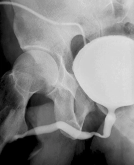
Background imageDiagnostic Collection: Voiding cystourethrogram, X-rays C018 / 0577

Voiding cystourethrogram, X-rays C018 / 0577
Voiding cystourethrogram. X-ray of a 54 year old male patient undergoing a voiding cystourethrogram. This procedure involves liquid containing a radio-opaque dye being placed in the bladder via a

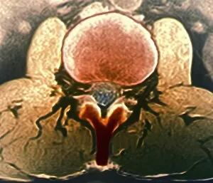
Background imageDiagnostic Collection: Healthy lumbar spine, MRI C018 / 0563

Healthy lumbar spine, MRI C018 / 0563
Healthy lumbar spine. Coloured magnetic resonance imaging (MRI) scan through the disc (red circle) between the second and third lumbar (lower back) vertebrae of a 30 year old patient

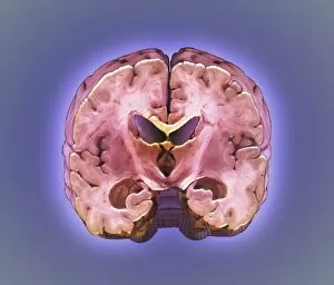
Background imageDiagnostic Collection: Healthy brain, MRI scan C018 / 0431

Healthy brain, MRI scan C018 / 0431
Healthy brain. Coloured 3D magnetic resonance imaging (MRI) scan of the healthy brain (excluding the brainstem) of a 37 year old. The cavities are the ventricles

Background imageDiagnostic Collection: Healthy brain, MRI scan C018 / 0430

Healthy brain, MRI scan C018 / 0430
Healthy brain. Coloured 3D magnetic resonance imaging (MRI) scan of the healthy brain (excluding the brainstem) of a 37 year old. The cavities are the ventricles

Background imageDiagnostic Collection: Uterine fibroids, MRI scan C018 / 0468

Uterine fibroids, MRI scan C018 / 0468
Uterine fibroids. Coloured magnetic resonance imaging (MRI) scan of the pelvis of a 47 year old female with two large fibroids (pink) in her uterus

Background imageDiagnostic Collection: Knee injury, 3D CT scan C018 / 0648

Knee injury, 3D CT scan C018 / 0648
Knee injury. Coloured 3D computed tomography (CT) scan of a knee with a meniscal tear (white line at right between bones)

Background imageDiagnostic Collection: Voiding cystourethrogram, X-rays C018 / 0578

Voiding cystourethrogram, X-rays C018 / 0578
Voiding cystourethrogram. Coloured X-ray of a 54 year old male patient undergoing a voiding cystourethrogram. This procedure involves liquid containing a radio-opaque dye being placed in the bladder

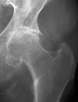
Background imageDiagnostic Collection: Osteoarthritis of the hip, X-ray C018 / 0593

Osteoarthritis of the hip, X-ray C018 / 0593
Osteoarthritis of the hip. X-ray the left hip of a 74 year old patient showing osteoarthritis. The ball at the top of the femur (thigh bone) fits into the socket of the hip

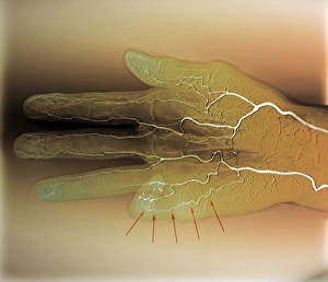
Background imageDiagnostic Collection: Blood supply of the hand, angiogram C018 / 0387

Blood supply of the hand, angiogram C018 / 0387
Blood supply of the hand. Coloured angiogram (X-ray) of the blood vessels of the right hand of a 65 year old patient with Dupuytrens contracture

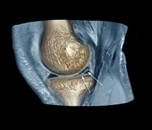
Background imageDiagnostic Collection: Knee injury, 3D CT scan C018 / 0639

Knee injury, 3D CT scan C018 / 0639
Knee injury. Coloured 3D computed tomography (CT) scan of a knee with a meniscal tear (indicated by arrows). The menisci are fibrocartilage crescents that act as shock absorbers between the femur

Background imageDiagnostic Collection: Hydrocephalus, CT scan C018 / 0568

Hydrocephalus, CT scan C018 / 0568
Hydrocephalus. Coloured computed tomography (CT) scan of the brain of a 22 year old patient with hydrocephalus (fluid in the brain)

Background imageDiagnostic Collection: Healthy brain, MRI scan C018 / 0429

Healthy brain, MRI scan C018 / 0429
Healthy brain. Coloured 3D magnetic resonance imaging (MRI) scan of the healthy brain (excluding the brainstem) of a 37 year old. The cavities are the ventricles

Background imageDiagnostic Collection: Fractured skull, X-ray C018 / 0591

Fractured skull, X-ray C018 / 0591
Fractured skull, X-ray. The fracture is the grey line at right that splits into two near the temple
Background imageDiagnostic Collection: Jeune syndrome, X-ray C018 / 0410

Jeune syndrome, X-ray C018 / 0410
Jeune syndrome. X-ray of the chest of a 2 year old with Jeune syndrome, also known as asphyxiating thoracic dystrophy. This inherited disease affects the development of the bones and cartilage
Background imageDiagnostic Collection: Cerebrotendinous xanthomatosis, MRI scan C018 / 0504

Cerebrotendinous xanthomatosis, MRI scan C018 / 0504
Cerebrotendinous xanthomatosis. Coloured magnetic resonance imaging (MRI) scan of the brain of a 46 year old patient with cerebrotendinous xanthomatosis (CTX)
Background imageDiagnostic Collection: Healthy lumbar spine, MRI C018 / 0562

Healthy lumbar spine, MRI C018 / 0562
Healthy lumbar spine. Coloured magnetic resonance imaging (MRI) scan through the disc (purple) between the second and third lumbar (lower back) vertebrae of a 30 year old patient

Background imageDiagnostic Collection: Arthritic hands, X-ray C018 / 0534

Arthritic hands, X-ray C018 / 0534
Arthritic hands. Coloured X-ray of the hands of a 72 year old patient with rheumatoid arthritis. The wrist and finger joints have become inflamed and swollen

Background imageDiagnostic Collection: Thrombophlebitis, angiogram C018 / 0529

Thrombophlebitis, angiogram C018 / 0529
Thrombophlebitis. Coloured angiogram (X-ray) of the brain of a 28 year old female patient who had thrombophelibitis of the superior sagittal sinus (indicated by arrows)

Background imageDiagnostic Collection: Rectal cancer, MRI scans C018 / 0511

Rectal cancer, MRI scans C018 / 0511
Rectal cancer. Coloured magnetic resonance imaging (MRI) scans of the pelvis of a 65 year old patient with a malignant (cancerous) tumour (purple) in the rectum, the last part of the large intestine

Background imageDiagnostic Collection: Knee injury, 3D CT scan C018 / 0645

Knee injury, 3D CT scan C018 / 0645
Knee injury. Coloured 3D computed tomography (CT) scan of a knee with a meniscal tear (blue line at right between bones). The menisci are fibrocartilage crescents that act as shock absorbers between


All Professionally Made to Order for Quick Shipping

"Unlocking the Secrets of Health: Exploring the World Tools" Medieval Urine Wheel: From ancient times to modern medicine, urine analysis has been a diagnostic tool used to detect various health conditions. Eyesight Test Chart: A simple yet crucial diagnostic tool that helps determine visual acuity and identify potential eye problems. Broken Wrist Bone, X-ray C017 / 7187: X-rays provide a clear view of fractured bones, aiding in accurate diagnosis and guiding treatment plans. Panoramic Dental X-ray: This comprehensive imaging technique reveals dental issues like cavities, impacted teeth, or jaw abnormalities for effective treatment planning. Home Cholesterol Test Kit: Empowering individuals with easy-to-use kits to monitor cholesterol levels at home and take proactive steps towards heart health. Full Body Scan, MRI Scan: Non-invasive and highly detailed imaging technology that allows doctors to visualize internal organs and diagnose complex medical conditions accurately. Brain Anatomy, MRI Scan: Advanced brain imaging enables neurologists to study intricate structures within the brain for precise diagnoses of neurological disorders. ECGs of a Normal Heart Rate, Artwork: Electrocardiograms help assess heart function by recording electrical activity; this artwork showcases what a healthy heartbeat looks like on paper. Tuberculosis, X-ray: Chest x-rays play a vital role in diagnosing tuberculosis by detecting characteristic lung abnormalities associated with this infectious disease. Normal EEG Readout of the Brain's Alpha Waves: Electroencephalography records brain wave patterns helping neurologists evaluate brain function and diagnose epilepsy or sleep disorders effectively. Eye Examination:A thorough examination assessing vision sharpness (visual acuity), peripheral vision tests (visual field), color perception evaluation among others is essential for maintaining good eye health Fractured Ankle, X-Ray.