Fraud Blocker Skip to main content

Cytoplasm Collection (page 5)

"Cytoplasm: The Dynamic Hub of Cellular Activity" The cytoplasm, a bustling and vibrant region within the cell, plays a crucial role in various cellular processes

Background imageCytoplasm Collection: Conceptual image of lyssavirus

Conceptual image of lyssavirus. Lyssavirus is a genus of viruses belonging to the family Rhabdoviridae. This group of RNA viruses includes the rabies virus traditionally associated with the disease

Background imageCytoplasm Collection: Microscopic view of red blood cells flowing inside lungs

Microscopic view of red blood cells flowing inside lungs

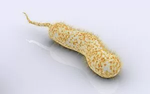
Background imageCytoplasm Collection: Conceptual image of vibrio cholerae causing cholera

Conceptual image of vibrio cholerae causing cholera

Background imageCytoplasm Collection: Artery cross section with red blood cell flow

Artery cross section with red blood cell flow

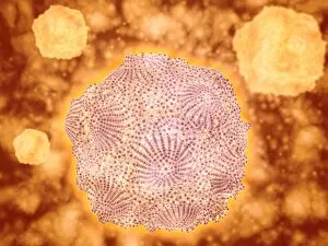
Background imageCytoplasm Collection: Microscopic view of Canine Parvovirus

Microscopic view of Canine Parvovirus. Canine Parvovirus is a highly contagious viral disease that is one of the most common causes of diarrhea in dogs under 6 months of age

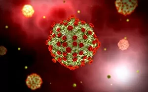
Background imageCytoplasm Collection: Microscopic view of HIV virus

Microscopic view of HIV virus

Background imageCytoplasm Collection: Surface of HIV infected microphage

Surface of HIV infected microphage
3D representation of the surface and interior of an HIV-infected macrophage obtained using newly developed tools for 3D imaging using ion-abrasion scanning electron microscopy

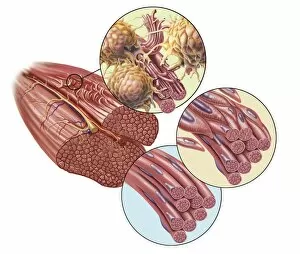
Background imageCytoplasm Collection: Torn muscle fibers with healing stages surrounding

Torn muscle fibers with healing stages surrounding. 1. Macrophage clean-up. 2. New cells migrate to repair. 3. Cells differentiate to become muscle cells

Background imageCytoplasm Collection: Red blood cells with white blood cells

Red blood cells with white blood cells

Background imageCytoplasm Collection: Influenza virus, glassy look

Influenza virus, glassy look

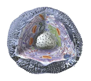
Background imageCytoplasm Collection: Model of animal cell, including cell nucleus, golgi body, lysosomes, centrioles, mitochondria

Model of animal cell, including cell nucleus, golgi body, lysosomes, centrioles, mitochondria, endoplasmic reticulum, ribosomes, cytoplasm, vesicles, thin plasma membrane

Background imageCytoplasm Collection: Human cell structure showing cytoplasm, cell membrane, nucleus, mitochondrion

Human cell structure showing cytoplasm, cell membrane, nucleus, mitochondrion

Background imageCytoplasm Collection: Eosinophil white blood cell, TEM C014 / 1438

Eosinophil white blood cell, TEM C014 / 1438
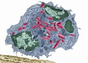
Eosinophil white blood cell. Transmission electron micrograph (TEM) of a section through an eosinophil. Eosinophils, like all white blood cells, are part of the bodys immune system

Background imageCytoplasm Collection: Eosinophil white blood cell, TEM C014 / 1437

Eosinophil white blood cell, TEM C014 / 1437
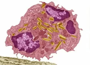
Eosinophil white blood cell. Transmission electron micrograph (TEM) of a section through an eosinophil. Eosinophils, like all white blood cells, are part of the bodys immune system

Background imageCytoplasm Collection: Eosinophil white blood cell, TEM C014 / 1439

Eosinophil white blood cell, TEM C014 / 1439
Eosinophil white blood cell. Transmission electron micrograph (TEM) of a section through an eosinophil. Eosinophils, like all white blood cells, are part of the bodys immune system

Background imageCytoplasm Collection: Golgi apparatus, artwork F006 / 9211

Golgi apparatus, artwork F006 / 9211
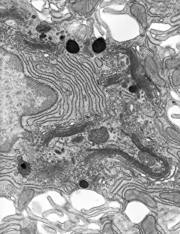
Computer artwork of the Golgi apparatus of the human cell. This organelle functions as a central delivery system for the cell

Background imageCytoplasm Collection: Golgi apparatus, artwork F006 / 9199

Golgi apparatus, artwork F006 / 9199
Computer artwork of the Golgi apparatus of the human cell. This organelle functions as a central delivery system for the cell

Background imageCytoplasm Collection: Golgi apparatus, artwork F006 / 9189

Golgi apparatus, artwork F006 / 9189
Computer artwork of the Golgi apparatus of the human cell. This organelle functions as a central delivery system for the cell

Background imageCytoplasm Collection: Enterocyte, TEM

Enterocyte, TEM
Enterocyte. Transmission electron micrograph (TEM) of a section through the cytoplasm and part of the nucleus of an enterocyte cell located in a crypt of Lieberkuhn of the small intestine

Background imageCytoplasm Collection: Dividing cell, TEM

Dividing cell, TEM
Dividing cell. Transmission electron micrograph (TEM) of a section through a cell dividing through the process of mitosis

Background imageCytoplasm Collection: Prokaryote cell, artwork C016 / 6263

Prokaryote cell, artwork C016 / 6263
Prokaryote cell. Computer artwork showing the internal structure of a typical prokaryotic cell. Prokaryotes are organisms whose cells lack a membrane-bound nucleus (karyon)

Background imageCytoplasm Collection: Eukaryote cell, artwork C016 / 6260

Eukaryote cell, artwork C016 / 6260
Eukaryote cell. Computer artwork showing the internal structure of a typical eukaryotic cell. Eukaryotes are organisms whose cells contain a membrane-bound nucleus (karyon, orange)

Background imageCytoplasm Collection: Prokaryote and eukaryote cells, artwork C016 / 6259

Prokaryote and eukaryote cells, artwork C016 / 6259
Prokaryote and eukaryote cells. Computer artwork comparing typical prokaryotic (top) and eukaryotic (bottom) cells. Prokaryotes are organisms whose cells lack a membrane-bound nucleus (karyon)

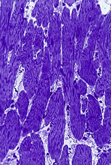
Background imageCytoplasm Collection: Heart muscle, light micrograph C016 / 0517

Heart muscle, light micrograph C016 / 0517
Heart muscle. Light micrograph of a section through heart (cardiac) muscle tissue, showing cardiomyocyte muscle cells. These cells have a centrally located nucleus and are branched

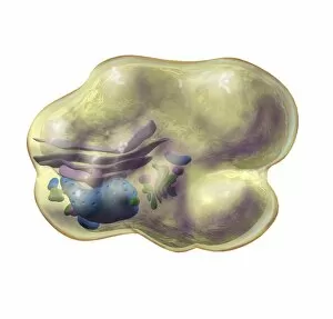
Background imageCytoplasm Collection: Fat cell anatomy, artwork

Fat cell anatomy, artwork
Fat cell anatomy. Artwork of an adipocyte (fat cell) and its internal organelles. The majority of the cell is filled with a lipid (fat) droplet
Background imageCytoplasm Collection: Golgi apparatus, artwork C015 / 6764

Golgi apparatus, artwork C015 / 6764
Computer artwork of the Golgi apparatus of the human cell. This organelle functions as a central delivery system for the cell

Background imageCytoplasm Collection: Myelinated nerve, TEM C016 / 5840

Myelinated nerve, TEM C016 / 5840
Myelinated nerve. Coloured transmission electron micrograph (TEM) of a section through a myelinated nerve fibre and Schwann cell

Background imageCytoplasm Collection: Myelinated nerve, TEM C016 / 5839

Myelinated nerve, TEM C016 / 5839
Myelinated nerve. Coloured transmission electron micrograph (TEM) of a section through a myelinated nerve fibre and Schwann cell

Background imageCytoplasm Collection: Myelinated nerve, TEM C016 / 5838

Myelinated nerve, TEM C016 / 5838
Myelinated nerve. Coloured transmission electron micrograph (TEM) of a section through a myelinated nerve fibre and Schwann cell

Background imageCytoplasm Collection: Myelinated nerve, TEM C016 / 5448

Myelinated nerve, TEM C016 / 5448
Myelinated nerve. Transmission electron micrograph (TEM) of a section through a myelinated nerve fibre and Schwann cell. Myelin (black)

Background imageCytoplasm Collection: Myelinated nerve, TEM C016 / 5370

Myelinated nerve, TEM C016 / 5370
Myelinated nerve. Transmission electron micrograph (TEM) of a section through a myelinated nerve fibre and Schwann cell (centre)

Background imageCytoplasm Collection: Animal cell, artwork C013 / 9985

Animal cell, artwork C013 / 9985
Animal cell. Computer artwork showing the cell organelles found inside a typical animal cell. The nucleus (large round) can be seen at centre

Background imageCytoplasm Collection: Animal cell, artwork C013 / 9984

Animal cell, artwork C013 / 9984
Animal cell. Computer artwork showing the cell organelles found inside a typical animal cell. The nucleus (large round) can be seen at centre

Background imageCytoplasm Collection: Myelinated nerves, SEM C013 / 7142

Myelinated nerves, SEM C013 / 7142
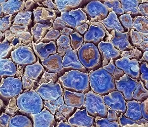
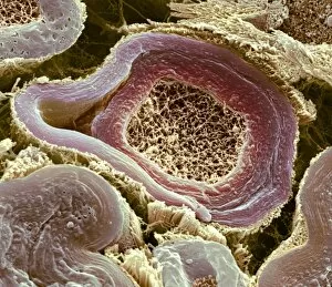
Myelinated nerves. Coloured scanning electron micrograph (SEM) of a section through the sciatic nerve, showing the myelinated nerve fibres (axons)

Background imageCytoplasm Collection: Myelinated nerves, SEM C013 / 7141

Myelinated nerves, SEM C013 / 7141
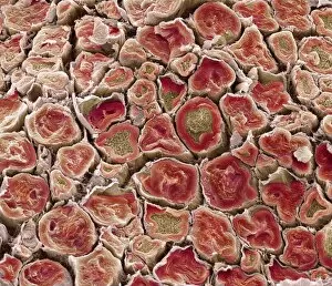
Myelinated nerves. Coloured scanning electron micrograph (SEM) of a section through the sciatic nerve, showing the myelinated nerve fibres (axons)

Background imageCytoplasm Collection: Myelinated nerves, SEM C013 / 7138

Myelinated nerves, SEM C013 / 7138
Myelinated nerves. Coloured scanning electron micrograph (SEM) of a section through a myelinated nerve fibre (axon, beige, centre) from the sciatic nerve

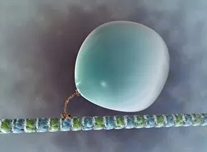
Background imageCytoplasm Collection: Intracellular transport, artwork C013 / 5001

Intracellular transport, artwork C013 / 5001
Intracellular transport. Computer artwork of a vesicle (sphere) being transported along a microtubule (blue and green) by a kinesin motor protein (orange)

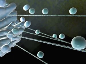
Background imageCytoplasm Collection: Intracellular transport, artwork C013 / 4997

Intracellular transport, artwork C013 / 4997
Intracellular transport. Computer artwork of vesicles (spheres) being transported from a Golgi body (blue, left) around the cell by microtubules (string-like)

Background imageCytoplasm Collection: Intracellular transport, artwork C013 / 4995

Intracellular transport, artwork C013 / 4995
Intracellular transport. Computer artwork of vesicles (spheres) being transported from a Golgi body (blue, top) around the cell by microtubules (string-like)

Background imageCytoplasm Collection: Cell from the epidermis of an onion

Cell from the epidermis of an onion
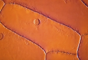
Light micrograph of a cell from the epidermis of an onion, showing the nucleus (round spot), cytoplasm and box-like cell wall. Magnification: x400 at 35mm size

Background imageCytoplasm Collection: Dendritic cell, TEM

Dendritic cell, TEM
Dendritic cell. Coloured transmission electron micrograph (TEM) of a section through dendritic cells. These are part of the human immune system

Background imageCytoplasm Collection: T-lymphocyte, TEM

T-lymphocyte, TEM
T-lymphocyte. Coloured transmission electron micrograph (TEM) of a section through a T- lymphocyte white blood cell. The cell contains a large nucleus (green/black)

Background imageCytoplasm Collection: HIV particles in infected cell, TEM

HIV particles in infected cell, TEM
HIV particles. Coloured transmission electron micrograph (TEM) of human immunodeficiency virus (HIV) particles (orange) in a host cell. HIV causes the disease AIDS

Background imageCytoplasm Collection: Nerve fibre node, TEM

Nerve fibre node, TEM
Nerve fibre node. Coloured transmission electron micrograph (TEM) of a cross-section through a nerve fibre (axon) at a node of Ranvier

Background imageCytoplasm Collection: Nerve fibres, TEM

Nerve fibres, TEM
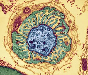
Nerve fibres. Coloured transmission electron micrograph (TEM) of a section through non- myelinated nerve fibres (blue). The fibres are arranged into bundles surrounded by the cytoplasm of Schwann

Background imageCytoplasm Collection: Immunofluorescent LM of an active macrophage

Immunofluorescent LM of an active macrophage
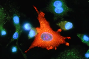
Active macrophage. Immunofluorescent light micrograph of an active macrophage white blood cell producing the enzyme inducible nitric oxide synthetase (iNOS, red)

Background imageCytoplasm Collection: Nerve demyelination, TEM

Nerve demyelination, TEM
Nerve demyelination. Coloured transmission electron micrograph (TEM) of a section through a Schwann cell and a nerve fibre, showing the early collapse of its myelin sheath

Background imageCytoplasm Collection: AIDS viruses budding from a cell, TEM

AIDS viruses budding from a cell, TEM
AIDS virus particles budding from the membrane of their host cell, coloured transmission electron micrograph (TEM). AIDS is a disease caused by the human immunodeficiency virus (HIV, red)


All Professionally Made to Order for Quick Shipping

"Cytoplasm: The Dynamic Hub of Cellular Activity" The cytoplasm, a bustling and vibrant region within the cell, plays a crucial role in various cellular processes. Through advanced techniques like Transmission Electron Microscopy (TEM) and fluorescent micrograph imaging, scientists have unraveled its intricate functions. In nerve cells, myelination of nerve fibers is a remarkable phenomenon that enhances signal transmission. TEM images reveal the mesmerizing beauty of these intricately wrapped fibers, highlighting the importance in maintaining efficient communication within our nervous system. During cell division, another captivating process captured through fluorescent micrographs, the cytoplasm orchestrates an elaborate dance to ensure accurate distribution of genetic material. These stunning visuals showcase how this dynamic organelle actively participates in life's fundamental cycle. Exploring further into TEM imagery unveils plasma cells with their distinctive appearance. Cytoplasmic structures are clearly visible as they produce antibodies vital for immune defense against pathogens such as human respiratory syncytial virus – depicted here under microscopic view. Fascinatingly, not only does cytoplasm support specialized cells but also aids viruses themselves. In AIDS infections visualized by TEM, we witness viral particles budding from host cells' cytoplasms – shedding light on the intricate relationship between pathogen and host. Delving deeper into cellular architecture reveals an illustration showcasing the nucleus containing essential components like mitochondria units, DNA strands coiled into chromosomes – all enveloped within a sea of cytoplasm. This conceptual image emphasizes its pivotal role in preserving genetic information and supporting vital cellular functions. Finally, a captivating artwork depicts platelets alongside red and white blood cells – symbolizing harmony within our circulatory system. The presence of these diverse cell types underscores how each relies on well-coordinated interactions facilitated by the ever-active cytoplasm. Whether it be myelination in nerve fibers or aiding viral replication or participating in cell division, the cytoplasm emerges as a dynamic and indispensable hub of cellular activity.