Fraud Blocker Skip to main content

Cellular Collection (page 8)

"Unveiling the Intricacies of Cellular Life: A Journey Through Micrographs" Budding yeast cell: Witness the birth of new life as a budding yeast cell emerges

Background imageCellular Collection: Chromatin remodelling factor and DNA C015 / 5155

Chromatin remodelling factor and DNA C015 / 5155
Chromatin remodelling factor and DNA, molecular model. The strands of DNA (deoxyribonucleic acid) are at left and right (pink-yellow and green-orange). This chromatin remodelling factor is ISW1a

Background imageCellular Collection: Importin protein complexed with enzyme C015 / 5149

Importin protein complexed with enzyme C015 / 5149
Importin protein complexed with enzyme, molecular model. Human importin13 (purple) complexed with SUMO-conjugating enzyme Ubc9 (red)

Background imageCellular Collection: Importin protein complexed with enzyme C015 / 5148

Importin protein complexed with enzyme C015 / 5148
Importin protein complexed with enzyme, molecular model. Human importin13 (green) complexed with SUMO-conjugating enzyme Ubc9 (pink)

Background imageCellular Collection: Sodium-potassium ion pump protein C016 / 2393

Sodium-potassium ion pump protein C016 / 2393
Sodium-potassium ion pump protein, molecular model. Sodium-potassium ATPase (adenosine triphosphatase) is an ATP-powered ion pump found in all animal cells

Background imageCellular Collection: Sodium-potassium ion pump protein C016 / 2392

Sodium-potassium ion pump protein C016 / 2392
Sodium-potassium ion pump protein, molecular model. Sodium-potassium ATPase (adenosine triphosphatase) is an ATP-powered ion pump found in all animal cells

Background imageCellular Collection: Sir3 gene silencer acting on DNA C016 / 2325

Sir3 gene silencer acting on DNA C016 / 2325
Sir3 gene silencer acting on DNA, molecular model. Sir3 (bright green) is acting on a circular strand of DNA (deoxyribonucleic acid, red and yellow)

Background imageCellular Collection: Sir3 gene silencer acting on DNA C016 / 2324

Sir3 gene silencer acting on DNA C016 / 2324
Sir3 gene silencer acting on DNA, molecular model. Sir3 (light blue) is acting on a circular strand of DNA (deoxyribonucleic acid, pink)

Background imageCellular Collection: HRas enzyme molecule C014 / 0010

HRas enzyme molecule C014 / 0010
HRas enzyme molecule. Molecular model showing the structure of a molecule of the enzyme GTPase HRas, also known as transforming protein p21
Background imageCellular Collection: NADH dehydrogenase molecule, artwork C014 / 0009

NADH dehydrogenase molecule, artwork C014 / 0009
NADH dehydrogenase molecule. Computer artwork showing the structure of a molecule of NADH dehydrogenase (NADH:ubiquinone reductase or Complex I)
Background imageCellular Collection: Mobile phone use C013 / 9870

Mobile phone use C013 / 9870
Mobile phone use. Close-up of a person at a train station, using a smartphone to check train times

Background imageCellular Collection: Mobile phone use C013 / 9869

Mobile phone use C013 / 9869
Mobile phone use. Close-up of a person viewing a map on a smartphone

Background imageCellular Collection: Oxoguanine glycosylase complex C013 / 8886

Oxoguanine glycosylase complex C013 / 8886
Oxoguanine glycosylase complex. Computer model showing a molecule of human aG DNA repair glycosylase (right) bound to an DNA molecule (left)

Background imageCellular Collection: Oxoguanine glycosylase complex C013 / 8884

Oxoguanine glycosylase complex C013 / 8884
Oxoguanine glycosylase complex. Computer model showing an 8-Oxoguanine glycosylase (OGG1) molecule (green) bound to a section of DNA (deoxyribonucleic acid, pink and blue)

Background imageCellular Collection: Microbiology laboratory C013 / 7251

Microbiology laboratory C013 / 7251
Microbiology laboratory. Microbiologist studying a sample under a microscope

Background imageCellular Collection: Microbiology laboratory C013 / 7250

Microbiology laboratory C013 / 7250
Microbiology laboratory. Microbiologist studying a sample under a microscope

Background imageCellular Collection: Microbiology laboratory C013 / 7249

Microbiology laboratory C013 / 7249
Microbiology laboratory. Microbiologist working on the production of vaccines

Background imageCellular Collection: Microbiology laboratory C013 / 7248

Microbiology laboratory C013 / 7248
Microbiology laboratory. Microbiologist working on the production of vaccines

Background imageCellular Collection: Microbiology laboratory C013 / 7247

Microbiology laboratory C013 / 7247
Microbiology laboratory. Microbiologist holding a Petri dish used to grow bacteria during vaccine research

Background imageCellular Collection: Microbiology laboratory C013 / 7246

Microbiology laboratory C013 / 7246
Microbiology laboratory. Microbiologist working on the production of vaccines

Background imageCellular Collection: Microbiology laboratory C013 / 7245

Microbiology laboratory C013 / 7245
Microbiology laboratory. Collection of glass bottles containing cultured bacterial samples in a microbiology lab

Background imageCellular Collection: Microbiology laboratory

Microbiology laboratory. Collection of glass bottles containing cultured bacterial samples in a microbiology lab

Background imageCellular Collection: Microbiology laboratory C013 / 7243

Microbiology laboratory C013 / 7243
Microbiology laboratory. Microbiologist working on the production of vaccines

Background imageCellular Collection: Microbiology laboratory C013 / 7241

Microbiology laboratory C013 / 7241
Microbiology laboratory. Microbiologist holding an electrophoresis gel. Gel electrophoresis uses an electric field applied across a gel matrix

Background imageCellular Collection: Microbiology laboratory C013 / 7242

Microbiology laboratory C013 / 7242
Microbiology laboratory. Microbiologist holding an electrophoresis gel. Gel electrophoresis uses an electric field applied across a gel matrix

Background imageCellular Collection: Microbiology laboratory C013 / 7240

Microbiology laboratory C013 / 7240
Microbiology laboratory. Microbiologist studying a sample under a microscope

Background imageCellular Collection: Microbiology laboratory C013 / 7239

Microbiology laboratory C013 / 7239
Microbiology laboratory. Microbiologist working on the production of vaccines

Background imageCellular Collection: Microbiology laboratory C013 / 7238

Microbiology laboratory C013 / 7238
Microbiology laboratory. Microbiologist working on the production of vaccines

Background imageCellular Collection: Microbiology laboratory C013 / 7237

Microbiology laboratory C013 / 7237
Microbiology laboratory. Microbiologist working on the production of vaccines

Background imageCellular Collection: Microbiology laboratory C013 / 7236

Microbiology laboratory C013 / 7236
Microbiology laboratory. Microbiologist working with a fermenter that is being used to grow bacterial cultures for analysis and production of vaccines

Background imageCellular Collection: Microbiology laboratory C013 / 7235

Microbiology laboratory C013 / 7235
Microbiology laboratory. Microbiologist working with a fermenter that is being used to grow bacterial cultures for analysis and production of vaccines

Background imageCellular Collection: Vitamin B12 transport protein C013 / 7184

Vitamin B12 transport protein C013 / 7184
Vitamin B12 transport protein, molecular model showing secondary structure. This receptor protein, known as BTUB, is from the Escherichia coli bacterium

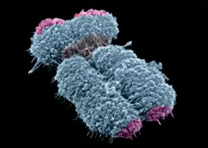
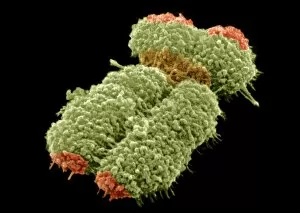
Background imageCellular Collection: Human chromosome, SEM C013 / 4999

Human chromosome, SEM C013 / 4999
Human chromosome. Coloured scanning electron micrograph (SEM) of a human chromosome. Chromosomes are a packaged form of a cells genetic material DNA (deoxyribonucleic acid)

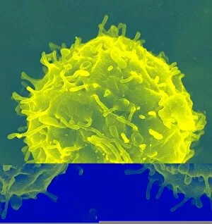
Background imageCellular Collection: Haematopoietic stem cell, SEM C013 / 5008

Haematopoietic stem cell, SEM C013 / 5008
Haematopoietic stem cell, coloured scanning electron micrograph (SEM). Stem cells can differentiate into any other cell type

Background imageCellular Collection: Haematopoietic stem cell, SEM C013 / 5007

Haematopoietic stem cell, SEM C013 / 5007
Haematopoietic stem cell, coloured scanning electron micrograph (SEM). Stem cells can differentiate into any other cell type

Background imageCellular Collection: Human chromosomes, SEM C013 / 5005

Human chromosomes, SEM C013 / 5005
Human chromosomes. Coloured scanning electron micrograph (SEM) of human chromosomes prepared with the harlequin staining technique

Background imageCellular Collection: Haematopoietic stem cell, SEM C013 / 5006

Haematopoietic stem cell, SEM C013 / 5006
Haematopoietic stem cell, coloured scanning electron micrograph (SEM). Stem cells can differentiate into any other cell type

Background imageCellular Collection: Human chromosome, SEM C013 / 4998

Human chromosome, SEM C013 / 4998
Human chromosome. Coloured scanning electron micrograph (SEM) of a human chromosome. Chromosomes are a packaged form of a cells genetic material DNA (deoxyribonucleic acid)

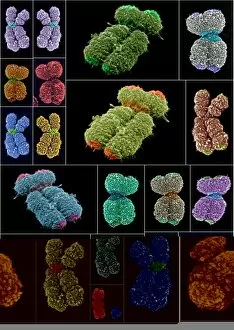
Background imageCellular Collection: Human chromosomes, SEMs

Human chromosomes, SEMs
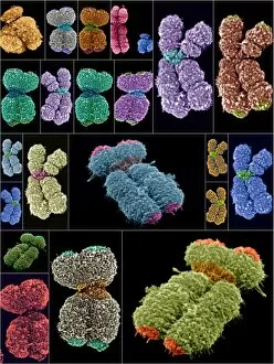
Human chromosomes. Collage of coloured scanning electron micrographs (SEMs) of human chromosomes. Chromosomes are a packaged form of a cells genetic material DNA (deoxyribonucleic acid)

Background imageCellular Collection: Human chromosomes, SEMs C013 / 4989

Human chromosomes, SEMs C013 / 4989
Human chromosomes. Collage of coloured scanning electron micrographs (SEMs) of human chromosomes. Chromosomes are a packaged form of a cells genetic material DNA (deoxyribonucleic acid)

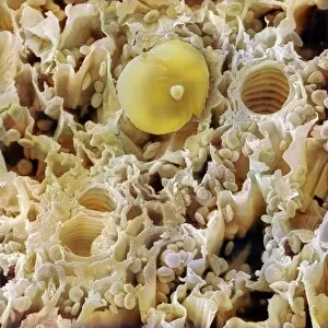
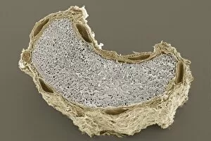
Background imageCellular Collection: Ginger rhizome, SEM

Ginger rhizome, SEM
Ginger rhizome. Coloured scanning electron micrograph (SEM) of a section through a rhizome (underground stem) from a ginger plant (Zingiber officinale)

Background imageCellular Collection: Cumin seed, SEM

Cumin seed, SEM
Cumin seed. Coloured scanning electron micrograph (SEM) of a cross-section through a seed from a cumin plant (Cuminum cyminum)

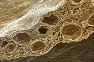
Background imageCellular Collection: Clove flower petal, SEM

Clove flower petal, SEM
Clove flower petal. Coloured scanning electron micrograph (SEM) of a cross-section through a clove flower petal. Cloves are the aromatic dried flower buds of the tropical tree Eugenia caryophyllata

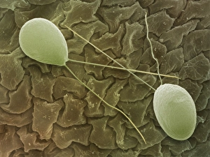
Background imageCellular Collection: Chlamydomonas sp. algae, SEM

Chlamydomonas sp. algae, SEM
Green algae. Coloured scanning electron micrograph (SEM) of two Chlamydomonas sp. green algae. These single-celled organisms are aquatic, living in freshwater habitats

Background imageCellular Collection: Dividing cell, light micrograph

Dividing cell, light micrograph
Cell division. Fluorescent light micrograph of a cell that has divided by mitosis, the asexual replication of a cell into two new cells

Background imageCellular Collection: Liver cells, light micrograph

Liver cells, light micrograph
Liver cells. Light micrograph of a section through a human liver. The liver cells are pink with purple nuclei. A duct canal and its surrounding tissue can be seen at lower left

Background imageCellular Collection: Intestinal cells, light micrograph

Intestinal cells, light micrograph
Intestinal cells. Light micrograph of a section through two folds (villi) in the small intestine. The villi are aligned vertically, with one at left and one at right

Background imageCellular Collection: Lung lining, SEM

Lung lining, SEM
Lung lining. Coloured scanning electron micrograph (SEM) of mucus-producing cells (orange, round) and cilia (yellow) lining a bronchus (lung airway)

Background imageCellular Collection: Large intestine, TEM

Large intestine, TEM
Large intestine. Coloured transmission electron micrograph (TEM) of a section through the wall of the large intestine. The surface consists of many small hair-like absorptive cells (green)


All Professionally Made to Order for Quick Shipping

"Unveiling the Intricacies of Cellular Life: A Journey Through Micrographs" Budding yeast cell: Witness the birth of new life as a budding yeast cell emerges, paving the way for future generations. Nerve and glial cells, light micrograph: Delve into the intricate network of nerve and glial cells that form the foundation of our nervous system. Dicotyledon plant stem, light micrograph: Explore the vibrant world within a dicotyledon plant stem, where cells work tirelessly to support growth and development. Rough endoplasmic reticulum, TEM: Peer into the fascinating realm of rough endoplasmic reticulum through a transmission electron microscope (TEM), revealing its role in protein synthesis. Dendritic cells, artwork: Marvel at an artistic representation showcasing dendritic cells' unique shape and function in immune responses. Cell structure: Uncover the hidden beauty within cellular structures as we unravel their complexity and significance in various biological processes. Mitosis, light micrograph: Observe nature's remarkable spectacle – mitosis – captured under a light microscope; witness how one cell divides into two with precision and grace. Cell division, fluorescent micrograph: Immerse yourself in a mesmerizing display of fluorescent colors capturing another facet of cell division's captivating dance. Dohle bodies in blood cell, micrograph: Discover peculiar "Dohle bodies" within blood cells through high-resolution microscopy—a clue to certain health conditions requiring further investigation. Acute promyelocytic leukemia, micrograph : Gain insight into acute promyelocytic leukemia by examining abnormal cellular characteristics observed under powerful magnification—an invaluable tool for diagnosis and treatment strategies Phagocytosis of fungal spores SEM : Witness nature's defense mechanism unfold as fungal spores are engulfed by cells in a captivating scanning electron microscope (SEM) image.