Fraud Blocker Skip to main content

Cellular Collection (page 3)

"Unveiling the Intricacies of Cellular Life: A Journey Through Micrographs" Budding yeast cell: Witness the birth of new life as a budding yeast cell emerges

Background imageCellular Collection: Mammatus lit up by lightning as it crosses a highway

Mammatus lit up by lightning as it crosses a highway
Mammatus from a collapsing severe thunderstorm is lit up by lightning as it crosses the highway; Durham, Oklahoma, United States of America

Background imageCellular Collection: A Man Stands In The Ocean With Items From Work And Vacation Flying Over His Head; Tarifa, Cadiz

A Man Stands In The Ocean With Items From Work And Vacation Flying Over His Head; Tarifa, Cadiz, Andalusia, Spain

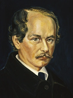
Background imageCellular Collection: SCHLEIDEN, Matthias Jakob (1804-1881). German botanist

SCHLEIDEN, Matthias Jakob (1804-1881). German botanist and co-founder of the cell theory. Oil

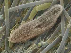
Background imageCellular Collection: Paramecium sp. protozoan, SEM

Paramecium sp. protozoan, SEM
Paramecium protozoan. Coloured scanning electron micrograph (SEM) of a Paramecium sp. protozoan. This single-celled organism is aquatic, living in freshwater habitats

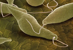
Background imageCellular Collection: Euglena gracilis, SEM

Euglena gracilis, SEM
Euglena gracilis protists. Coloured scanning electron micrograph (SEM) of Euglena gracilis protists. These organisms possess a unique combination of plant and animal characters. Like the algae, E

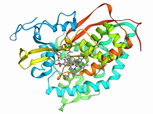
Background imageCellular Collection: Cytochrome P450 complex F006 / 9669

Cytochrome P450 complex F006 / 9669
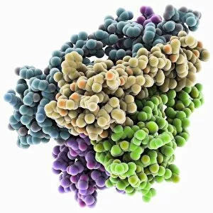
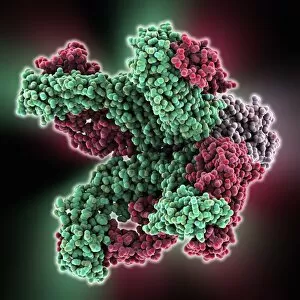
Cytochrome P450 complex. Molecular model of a complex composed of cytochrome P450, carbon monoxide and camphor. Cytochrome molecules perform oxidation and reduction reactions for electron transport

Background imageCellular Collection: Myeloblast blood cell, light micrograph

Myeloblast blood cell, light micrograph
Myeloblast blood cell. Light micrograph of blood cells, including a myeloblast, a precursor for a type of white blood cell (leucocyte). Myeloblasts differentiate into granulocytes

Background imageCellular Collection: Promyelocyte blood cell, light micrograph

Promyelocyte blood cell, light micrograph. This blood cell (centre) is a precursor for a type of white blood cell called a granulocyte, formed by granulopoiesis in the bone marrow

Background imageCellular Collection: Chronic lymphocytic leukaemia, micrograph

Chronic lymphocytic leukaemia, micrograph
Chronic lymphocytic leukaemia. Light micrograph of blood cells sampled from a lymphatic ganglion in a case of chronic lymphocytic leukaemia, also called chronic lymphoid leukaemia (CLL)

Background imageCellular Collection: Macrophage and lymphocytes, TEM

Macrophage and lymphocytes, TEM
Macrophage and lymphocytes, transmission electron micrograph (TEM). Macrophage surrounded by lymphocytes in a lymph node. This view includes many intercellular contacts

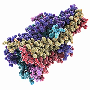
Background imageCellular Collection: MscL ion channel protein structure

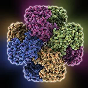
MscL ion channel protein structure. Molecular model showing the protein structure of a Mechanosensitive Channel of Large Conductance (MscL) from a Mycobacterium tuberculosis bacterium

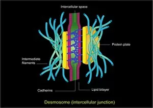
Background imageCellular Collection: Desmosome cell junction, artwork

Desmosome cell junction, artwork
Desmosome cell junction. Computer artwork showing the structure of an adhesion junction, or desmosome. Desmosomes form the most common type of junction between epithelial cells

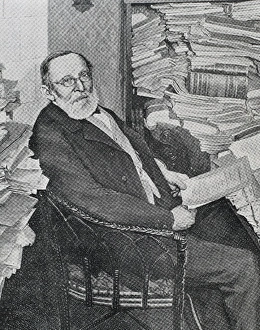
Background imageCellular Collection: VIRCHOW, Rudolf (1821-1902). German doctor, anthropologist

VIRCHOW, Rudolf (1821-1902). German doctor, anthropologist, pathologist, prehistorian, biologist and politician, known for his advancement of public health. Engraving

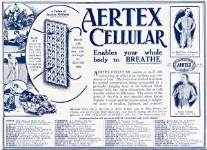
Background imageCellular Collection: Advert for Aertex Cellular mens undergarments 1912

Advert for Aertex Cellular mens undergarments 1912
Enables your whole body to BREATH. Non-conductor of heat, allowing the body to be clothed in lightweight Aertex Cellular and maintain its normal temperture. 1912

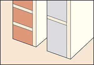
Background imageCellular Collection: Digital Illustration of solid masonry and cellular block wall material

Digital Illustration of solid masonry and cellular block wall material

Background imageCellular Collection: Business man outside a bank

Business man outside a bank

Background imageCellular Collection: Business man greeting someone outside a bank

Business man greeting someone outside a bank

Background imageCellular Collection: Young woman speaking on her mobile phone, smiling

Young woman speaking on her mobile phone, smiling

Background imageCellular Collection: Young woman speaking on her mobile phone

Young woman speaking on her mobile phone

Background imageCellular Collection: Further evolution of the cellular kite

Further evolution of the cellular kite. Hargrave Slide 36

Background imageCellular Collection: Hookes observations of cellular structure of cork (fig 1) and sprig of Sensible

Hookes observations of cellular structure of cork (fig 1) and sprig of Sensible (Sensitive) plant (fig 2). First use of word cell to name honeycomb nature of cork

Background imageCellular Collection: Farmer talking on mobile phone, standing beside tractor and trailer with straw bales, Sweden

Farmer talking on mobile phone, standing beside tractor and trailer with straw bales, Sweden

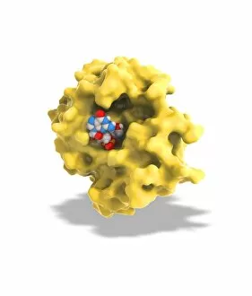
Background imageCellular Collection: Ricin A-chain, artwork C017 / 3653

Ricin A-chain, artwork C017 / 3653
Ricin A-chain. Computer artwork showing the enzymatically active A-chain from a molecule of the toxic protein ricin. Ricin comprises two entwined amino acid chains; A (seen here) and B (not shown)

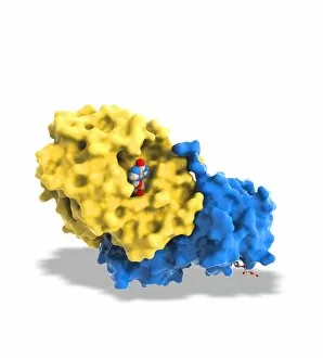
Background imageCellular Collection: Ricin molecule, artwork C017 / 3652

Ricin molecule, artwork C017 / 3652
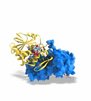
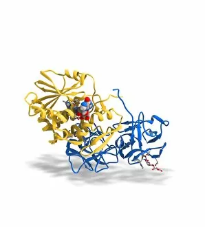
Ricin molecule. Computer artwork showing the structure of a molecule of the toxic protein ricin. Ricin comprises two entwined amino acid chains; A (yellow) and B (blue)

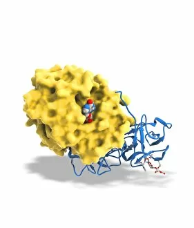
Background imageCellular Collection: Ricin molecule, artwork C017 / 3651

Ricin molecule, artwork C017 / 3651
Ricin molecule. Computer artwork showing the structure of a molecule of the toxic protein ricin. Ricin comprises two entwined amino acid chains; A (yellow) and B (blue)

Background imageCellular Collection: Ricin molecule, artwork C017 / 3650

Ricin molecule, artwork C017 / 3650
Ricin molecule. Computer artwork showing the structure of a molecule of the toxic protein ricin. Ricin comprises two entwined amino acid chains; A (yellow) and B (blue)

Background imageCellular Collection: Photosynthesis, artwork C017 / 0788

Photosynthesis, artwork C017 / 0788
Photosynthesis. Conceptual computer artwork of photosynthesis in a plant cell. Photosynthesis is the process by which most plants convert sunlight (coming from top left) into chemical energy

Background imageCellular Collection: Bacteria shapes, artwork F007 / 9891

Bacteria shapes, artwork F007 / 9891
Computer artwork showing the three basic forms of bacteria: in the front (red) spirochetes), in the middle (green) bacilli and in the background (blue) cocci

Background imageCellular Collection: Human chromosome

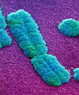
Human chromosome. Coloured scanning electron micrograph (SEM) of a human chromosome. Chromosomes occur in the nucleus of every cell in the body

Background imageCellular Collection: Cells, conceptual artwork

Cells, conceptual artwork
Cells, conceptual computer artwork

Background imageCellular Collection: Breast cancer, light micrograph F005 / 6079

Breast cancer, light micrograph F005 / 6079
Breast cancer. Light micrograph of a section through a ductal carcinoma (cancer) of the breast. This is a cancer that arises from the lining of the milk ducts

Background imageCellular Collection: Secondary liver cancer, light micrograph F005 / 6100

Secondary liver cancer, light micrograph F005 / 6100
Secondary liver cancer. Light micrograph of a section through liver tissue showing a secondary cancer that had metastasised (spread) from a primary small cell lung cancer

Background imageCellular Collection: Nerve cells, artwork F005 / 0626

Nerve cells, artwork F005 / 0626
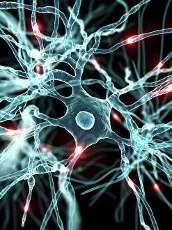
Nerve cells, computer artwork

Background imageCellular Collection: Breast cancer, light micrograph F005 / 6080

Breast cancer, light micrograph F005 / 6080
Breast cancer. Light micrograph of a section through a ductal carcinoma (cancer) of the breast. This is a cancer that arises from the lining of the milk ducts

Background imageCellular Collection: Bacterium, artwork

Bacterium, artwork
Bacterium, computer artwork

Background imageCellular Collection: Multicellular organism, conceptual image

Multicellular organism, conceptual image
Multicellular organism, conceptual computer artwork

Background imageCellular Collection: Nerve cells, artwork F005 / 0583

Nerve cells, artwork F005 / 0583
Nerve cells, computer artwork

Background imageCellular Collection: Nerve cells, artwork F005 / 0582

Nerve cells, artwork F005 / 0582
Nerve cells, computer artwork

Background imageCellular Collection: Nerve cells, artwork F005 / 0584

Nerve cells, artwork F005 / 0584
Nerve cells, computer artwork

Background imageCellular Collection: Potassium ion channel protein structure

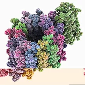
Potassium ion channel protein structure. Molecular model of a KcsA potassium ion (K+) channel from Streptomyces lividans bacteria

Background imageCellular Collection: Potassium ion channel beta subunit

Potassium ion channel beta subunit. Molecular model showing the structure a beta subunit of a voltage-dependent potassium (K+) channel

Background imageCellular Collection: KCNQ ion channel protein structure

KCNQ ion channel protein structure. Molecular model showing the protein structure of an ion channel domain. Ion channels are membrane-spanning proteins that form a pathway for the movement of

Background imageCellular Collection: Potassium ion channel cavity structure

Potassium ion channel cavity structure. Molecular model showing the structure of a cavity formed by potassium ion channel proteins

Background imageCellular Collection: ATP synthase molecule C014 / 0880

ATP synthase molecule C014 / 0880
ATP synthase molecule. Molecular model showing the structure of ATP synthase (ATPase) subunit C. ATPase is an important enzyme that provides energy for cells through the synthesis of adenosine

Background imageCellular Collection: Ricin A-chain, artwork C017 / 3654

Ricin A-chain, artwork C017 / 3654
Ricin A-chain. Computer artwork showing the enzymatically active A-chain from a molecule of the toxic protein ricin. Ricin comprises two entwined amino acid chains; A (seen here) and B (not shown)

Background imageCellular Collection: Nucleus and endoplasmic reticulum F006 / 9196

Nucleus and endoplasmic reticulum F006 / 9196
Computer artwork showing part of a human or eukaryotic cell. In the middle the nucleus which has a membrane with nuclear pores. Inside the nucleus is the DNA

Background imageCellular Collection: Ricin molecule, artwork C017 / 3649

Ricin molecule, artwork C017 / 3649
Ricin molecule. Computer artwork showing the structure of a molecule of the toxic protein ricin. Ricin comprises two entwined amino acid chains; A (yellow) and B (blue)

Background imageCellular Collection: Cytochrome C, molecular model F006 / 9696

Cytochrome C, molecular model F006 / 9696
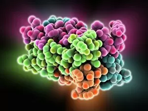
Cytochrome C, molecular model. Cytochrome molecules perform oxidation and reduction reactions for electron transport, a chain of reactions used to power cellular processes that require energy


All Professionally Made to Order for Quick Shipping

"Unveiling the Intricacies of Cellular Life: A Journey Through Micrographs" Budding yeast cell: Witness the birth of new life as a budding yeast cell emerges, paving the way for future generations. Nerve and glial cells, light micrograph: Delve into the intricate network of nerve and glial cells that form the foundation of our nervous system. Dicotyledon plant stem, light micrograph: Explore the vibrant world within a dicotyledon plant stem, where cells work tirelessly to support growth and development. Rough endoplasmic reticulum, TEM: Peer into the fascinating realm of rough endoplasmic reticulum through a transmission electron microscope (TEM), revealing its role in protein synthesis. Dendritic cells, artwork: Marvel at an artistic representation showcasing dendritic cells' unique shape and function in immune responses. Cell structure: Uncover the hidden beauty within cellular structures as we unravel their complexity and significance in various biological processes. Mitosis, light micrograph: Observe nature's remarkable spectacle – mitosis – captured under a light microscope; witness how one cell divides into two with precision and grace. Cell division, fluorescent micrograph: Immerse yourself in a mesmerizing display of fluorescent colors capturing another facet of cell division's captivating dance. Dohle bodies in blood cell, micrograph: Discover peculiar "Dohle bodies" within blood cells through high-resolution microscopy—a clue to certain health conditions requiring further investigation. Acute promyelocytic leukemia, micrograph : Gain insight into acute promyelocytic leukemia by examining abnormal cellular characteristics observed under powerful magnification—an invaluable tool for diagnosis and treatment strategies Phagocytosis of fungal spores SEM : Witness nature's defense mechanism unfold as fungal spores are engulfed by cells in a captivating scanning electron microscope (SEM) image.