Fraud Blocker Skip to main content

Cell Biology Collection (page 17)

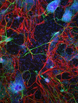
"Exploring the Intricacies of Cell Biology: A Fascinating Journey into Cellular Life" Nerve and glial cells, captured through a mesmerizing light micrograph

Background imageCell Biology Collection: Cytoskeleton in unicellular parasite, SEM C018 / 0518

Cytoskeleton in unicellular parasite, SEM C018 / 0518
Cytoskeleton in unicellular parasite, coloured scanning electron micrograph (SEM). All cells have a support and transport network called the cytoskeleton

Background imageCell Biology Collection: Gene expression, conceptual illustration C018 / 0746

Gene expression, conceptual illustration C018 / 0746
Gene expression, conceptual illustration. Every cell in an organism contains every single gene that makes up the organisms genome. However, they are not all active (expressed) in each cell

Background imageCell Biology Collection: Animal cell, illustration C018 / 0734

Animal cell, illustration C018 / 0734
Animal cell. Illustration showing the organelles present in an animal cell. At lower centre is the nucleus, which contains the cells genetic information in the form of DNA (deoxyribonucleic acid)

Background imageCell Biology Collection: Mechanical stress on a cell, illustration C018 / 0776

Mechanical stress on a cell, illustration C018 / 0776
Mechanical stress on a cell, conceptual illustration. Mechanical stress can have an effect on a number of processes within a cell, including polarity, differentiation, growth and migration

Background imageCell Biology Collection: Cellular polarity, illustration C018 / 0775

Cellular polarity, illustration C018 / 0775
Cellular Polarity. Conceptual illustration of cell polarity research and its medical implications

Background imageCell Biology Collection: Microtubule formation, illustration C018 / 0804

Microtubule formation, illustration C018 / 0804
Microtubule formation, illustration. Microtubules are polymers of the protein tubulin and are a component of the cytoskeleton

Background imageCell Biology Collection: Migrating cell, illustration C018 / 0754

Migrating cell, illustration C018 / 0754
Migrating cell. Time-lapse illustration of a migrating cell, using outlines to show the previous cell boundaries. Cell migration is key to metastasis (spread) and the progression of cancer

Background imageCell Biology Collection: Eye muscle, TEM C014 / 1467

Eye muscle, TEM C014 / 1467
Eye muscle. Transmission electron micrograph (TEM) of a section through a striated muscle cell from the ciliary muscle of a human eye

Background imageCell Biology Collection: Intestinal gland cell, TEM C014 / 1442

Intestinal gland cell, TEM C014 / 1442
Intestinal gland cell. Transmission electron micrograph (TEM) of a section through an undifferentiated cell from the intestinal tract, showing extensive rough endoplasmic reticulum (ER)

Background imageCell Biology Collection: Mouth cancer cell, SEM C014 / 1460

Mouth cancer cell, SEM C014 / 1460
Mouth cancer cell. Coloured scanning electron micrograph (SEM) of a squamous cell carcinoma (cancer) cell from a human mouth

Background imageCell Biology Collection: Genetic metaphase analysis C019 / 0289

Genetic metaphase analysis C019 / 0289
Genetic metaphase analysis. Close-up of a microscope being used in a genetics laboratory to analyse the metaphase stage of cell division

Background imageCell Biology Collection: Genetic metaphase analysis C019 / 0288

Genetic metaphase analysis C019 / 0288
Genetic metaphase analysis. Equipment used in a genetics laboratory to analyse the metaphase stage of cell division. During this stage

Background imageCell Biology Collection: Eye muscle, TEM C014 / 1466

Eye muscle, TEM C014 / 1466
Eye muscle. Transmission electron micrograph (TEM) of a section through a striated muscle cell from the ciliary muscle of a human eye

Background imageCell Biology Collection: Intestinal microvilli, SEM C014 / 1452

Intestinal microvilli, SEM C014 / 1452
Intestinal microvilli. Coloured transmission electron micrograph (SEM) of a transverse section through microvilli, showing their interiors

Background imageCell Biology Collection: Mouth cancer cell, SEM C014 / 1459

Mouth cancer cell, SEM C014 / 1459
Mouth cancer cell. Coloured scanning electron micrograph (SEM) of a squamous cell carcinoma (cancer) cell from a human mouth

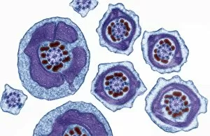
Background imageCell Biology Collection: Sperm tails, TEM C014 / 1463

Sperm tails, TEM C014 / 1463
Sperm tails. Transmission electron micrograph (TEM) of sections through the tails of sperm (spermatozoa) cells at various levels

Background imageCell Biology Collection: Intestinal microvilli, SEM C014 / 1451

Intestinal microvilli, SEM C014 / 1451
Intestinal microvilli. Coloured transmission electron micrograph (SEM) of a transverse section through microvilli, showing their interiors

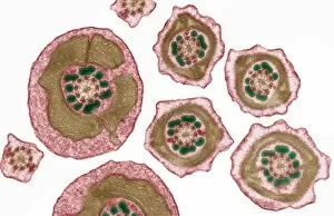
Background imageCell Biology Collection: Sperm tails, TEM C014 / 1465

Sperm tails, TEM C014 / 1465
Sperm tails. Transmission electron micrograph (TEM) of sections through the tails of sperm (spermatozoa) cells at various levels

Background imageCell Biology Collection: Intestinal microvilli, TEM C014 / 1454

Intestinal microvilli, TEM C014 / 1454
Intestinal microvilli. Transmission electron micrograph (TEM) of a section through an epithelial cell from a human small intestine, showing the densely packed microvilli

Background imageCell Biology Collection: Fat cells, SEM C014 / 0365

Fat cells, SEM C014 / 0365
Fat cells. Coloured scanning electron micrograph (SEM) of adipocytes (fat cells). Adipocytes store energy as an insulating layer of fat

Background imageCell Biology Collection: Intestinal microvilli, TEM C014 / 1453

Intestinal microvilli, TEM C014 / 1453
Intestinal microvilli. Transmission electron micrograph (TEM) of a section through an epithelial cell from a human small intestine, showing the densely packed microvilli

Background imageCell Biology Collection: Monocyte white blood cell, TEM

Monocyte white blood cell, TEM
Monocyte white blood cell. Transmission electron micrograph (TEM) of a section through a monocyte white blood cell, showing the cell nucleus (blue)

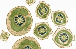
Background imageCell Biology Collection: Sperm tails, TEM C014 / 1464

Sperm tails, TEM C014 / 1464
Sperm tails. Transmission electron micrograph (TEM) of sections through the tails of sperm (spermatozoa) cells at various levels

Background imageCell Biology Collection: Zinc finger bound to DNA C014 / 0864

Zinc finger bound to DNA C014 / 0864
Zinc finger bound to DNA. Molecular model showing a zinc finger molecule bound to a strand of DNA (deoxyribonucleic acid)

Background imageCell Biology Collection: Epstein-Barr virus protein bound to DNA C014 / 0875

Epstein-Barr virus protein bound to DNA C014 / 0875
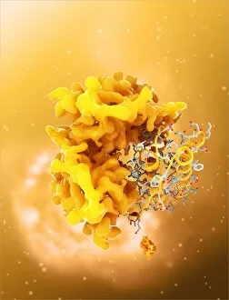
Epstein-Barr virus protein bound to DNA. Computer model showing a molecule of Epstein-Barr nuclear antigen 1 (EBNA1) bound to a strand of DNA (deoxyribonucleic acid)

Background imageCell Biology Collection: ATP synthase molecule

ATP synthase molecule. Molecular model showing the structure of ATP synthase (ATPase) subunit C. ATPase is an important enzyme that provides energy for cells through the synthesis of adenosine

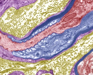
Background imageCell Biology Collection: Nerve fibre node, TEM C014 / 1458

Nerve fibre node, TEM C014 / 1458
Nerve fibre node. Transmission electron micrograph (TEM) of a longitudinal section through a nerve fibre at a node of Ranvier, showing the nerve tissue (red) and the myelin sheath (dark blue)

Background imageCell Biology Collection: Leptin molecule C014 / 4904

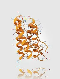
Leptin molecule C014 / 4904
Leptin molecule. Computer model showing the structure of a molecule of the hormone leptin. Leptin is produced by adipose (fat) tissue
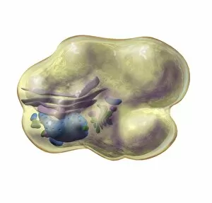
Background imageCell Biology Collection: Human tissue structure, historical model

Human tissue structure, historical model
Human tissue structure. 1960s anatomical model of a section through human body tissue, showing the arrangement and structure of the cells

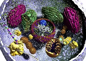
Background imageCell Biology Collection: Animal cell organelles, artwork

Animal cell organelles, artwork
Animal cell organelles. Artwork showing the organelles in a eukaryotic cell. This is an animal cell. Structures include the nucleus (centre) which has a membrane with nuclear pores (purple)

Background imageCell Biology Collection: Mitochondrion structure, artwork

Mitochondrion structure, artwork
Mitochondrion structure. Artwork showing the internal structure of a mitochondrion. This structure, found in eukaryotic cells, is the site of energy production

Background imageCell Biology Collection: Leptin molecule C014 / 4907

Leptin molecule C014 / 4907
Leptin molecule. Computer model showing the crystal structure of a molecule of the hormone leptin. Leptin is produced by adipose (fat) tissue

Background imageCell Biology Collection: Squamous epithelium C014 / 4643

Squamous epithelium C014 / 4643
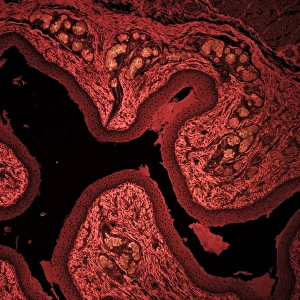
Squamous epithelium. Laser scanning confocal micrograph (LSCM) of a section through stratified squamous epithelial cells. Epithelial tissues line the cavities

Background imageCell Biology Collection: Squamous epithelium, confocal micrograph C014 / 4642

Squamous epithelium, confocal micrograph C014 / 4642
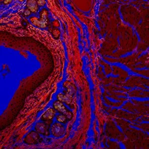
Squamous epithelium. Laser scanning confocal micrograph (LSCM) of a section through stratified squamous epithelial cells. Epithelial tissues line the cavities

Background imageCell Biology Collection: Mouth cancer cells, SEM

Mouth cancer cells, SEM
Mouth cancer cells. Coloured scanning electron micrograph (SEM) of squamous cell carcinoma (cancer) cells from a human mouth

Background imageCell Biology Collection: Fat cell anatomy, artwork

Fat cell anatomy, artwork
Fat cell anatomy. Artwork of an adipocyte (fat cell) and its internal organelles. The majority of the cell is filled with a lipid (fat) droplet

Background imageCell Biology Collection: Eukaryotic cell nucleus, artwork

Eukaryotic cell nucleus, artwork
Eukaryotic cell nucleus. Artwork of the internal structure and contents of the nucleus of a eukaryotic cell. The nucleus has been sectioned in half

Background imageCell Biology Collection: Mitochondrial structure, artwork

Mitochondrial structure, artwork
Mitochondrial structure. Computer artwork of a section through a mitochondrion, showing the internal structure and a loop of mitochondrial DNA (deoxyribonucleic acid, mtDNA)

Background imageCell Biology Collection: Leptin molecule and fat cells C014 / 4906

Leptin molecule and fat cells C014 / 4906
Leptin molecule and fat cells. Computer artwork showing a molecule of the hormone leptin with adipose (fat) cells (round), from which it is produced

Background imageCell Biology Collection: Mouth cancer cell dividing, SEM

Mouth cancer cell dividing, SEM
Mouth cancer dividing. Coloured scanning electron micrograph (SEM) of a squamous cell carcinoma (cancer) cell from a human mouth undergoing mitosis (nuclear division)

Background imageCell Biology Collection: Bacterial outer membrane protein molecule

Bacterial outer membrane protein molecule. Computer model showing a part of the secondary structure of a molecule of outer membrane protein A from Escherichia coli (E. coli) bacteria

Background imageCell Biology Collection: Mouth cancer cell, SEM

Mouth cancer cell, SEM
Mouth cancer cell. Coloured scanning electron micrograph (SEM) showing the surface of a squamous cell carcinoma (cancer) cell from a human mouth

Background imageCell Biology Collection: Induced stem cells, light micrograph

Induced stem cells, light micrograph
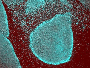
Induced stem cells. Light micrograph of a cultured colony of induced human pluripotent stem (IPS) cells. Oct-4 (octamer-binding transcription factor 4) is cyan

Background imageCell Biology Collection: Induced nerve stem cells, micrograph

Induced nerve stem cells, micrograph
Induced nerve stem cells. Fluorescence light micrograph of neural (nerve) stem cells that have been created (induced) from human adult skin fibroblast cells by gene manipulation

Background imageCell Biology Collection: Dividing liver cancer cell, SEM

Dividing liver cancer cell, SEM
Dividing liver cancer cell. Coloured scanning electron micrograph (SEM) of a hepatocellular carcinoma (HCC) cell undergoing mitosis (nuclear division)

Background imageCell Biology Collection: Stem cell-derived nerve cells

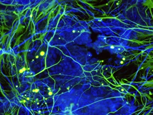
Stem cell-derived nerve cells. Fluorescence light micrograph of neural (nerve) stem cells that have been derived from human embryonic stem cells (HESC)


All Professionally Made to Order for Quick Shipping

"Exploring the Intricacies of Cell Biology: A Fascinating Journey into Cellular Life" Nerve and glial cells, captured through a mesmerizing light micrograph, reveal the intricate network that supports our nervous system. T lymphocytes battling cancer cells - witness this epic battle in stunning detail with SEM C001 / 1679 imagery. Anaesthetic inhibiting an ion channel - delve into the world of cellular communication as we uncover how anaesthetics affect these vital channels (C015 / 6718). Glial cells take center stage in a captivating confocal light micrograph, showcasing their crucial role in supporting neurons. Witness the beauty of glial stem cell culture under a microscope; it's like exploring an enchanting galaxy within our own bodies (light micrograph). Dendritic cells come to life through exquisite artwork, highlighting their pivotal role in immune response and defense mechanisms. Pine pollen grains unveil their intricate structure under a light microscope – nature's tiny marvels never cease to amaze us. Dive deep into the inner workings of pine stems with a captivating light micrograph – discover the building blocks that support these majestic trees. Explore the hidden wonders of lime tree stems through a mesmerizing light micrograph – witness nature's architectural masterpiece up close. Uncover various cell types brought to life through breathtaking artwork – each one playing its unique part in maintaining life's delicate balance. Immerse yourself in neural stem cell culture and witness firsthand how these remarkable cells hold immense potential for regenerative medicine. The artistic portrayal of cell membranes (C013 / 7467) invites you to ponder upon their crucial role as gatekeepers controlling molecular traffic within living organisms. Embark on this awe-inspiring journey into cell biology and unlock the secrets that shape all living beings from within.