Fraud Blocker Skip to main content

Blood Vessels Collection (page 9)

Blood vessels are the intricate network of highways that carry life-sustaining blood throughout our bodies

Background imageBlood Vessels Collection: Heart attack, conceptual artwork C013 / 5930

Heart attack, conceptual artwork C013 / 5930
Heart attack, conceptual computer artwork. A heart attack results from the interruption of blood supply to a part of the heart, causing heart cells to die

Background imageBlood Vessels Collection: Human anatomy, 1823 C017 / 8057

Human anatomy, 1823 C017 / 8057
Human anatomy. 19th-century illustrations showing the anatomy of a human skull (upper left), brain and spinal column (upper centre), chest and abdominal organs (lungs, heart, liver, stomach)

Background imageBlood Vessels Collection: Single-kidney anatomy, artwork C016 / 2902

Single-kidney anatomy, artwork C016 / 2902
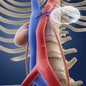
Single-kidney anatomy. Artwork showing an anterior (frontal) view of human kidneys in relation to the main torso blood vessels (vena cava, blue; aorta, red) and the male abdomen

Background imageBlood Vessels Collection: Single-kidney anatomy, artwork C016 / 2903

Single-kidney anatomy, artwork C016 / 2903
Single-kidney anatomy. Artwork showing a posterior (from behind) view of human kidneys in relation to the main torso blood vessels (vena cava, blue; aorta, red) and the male abdomen

Background imageBlood Vessels Collection: Single-kidney anatomy, artwork C016 / 2901

Single-kidney anatomy, artwork C016 / 2901
Single-kidney anatomy. Artwork showing an anterior (frontal) view of human kidneys in relation to the main torso blood vessels (vena cava, blue; aorta, red) and bones of the male skeleton

Background imageBlood Vessels Collection: Single-kidney anatomy, artwork C016 / 2898

Single-kidney anatomy, artwork C016 / 2898
Single-kidney anatomy. Artwork showing an oblique right-frontal view of human kidneys in relation to the main torso blood vessels (vena cava, blue; aorta)

Background imageBlood Vessels Collection: Single-kidney anatomy, artwork C016 / 2897

Single-kidney anatomy, artwork C016 / 2897
Single-kidney anatomy. Artwork showing a posterior (from behind) view of human kidneys in relation to the bones of the skeleton. One kidney (brown, upper right) has a normal appearance

Background imageBlood Vessels Collection: Single-kidney anatomy, artwork C016 / 2900

Single-kidney anatomy, artwork C016 / 2900
Single-kidney anatomy. Artwork showing an anterior (frontal) view of human kidneys in relation to the main torso blood vessels (vena cava, blue; aorta, red) and bones of the skeleton

Background imageBlood Vessels Collection: Single-kidney anatomy, artwork C016 / 2896

Single-kidney anatomy, artwork C016 / 2896
Single-kidney anatomy. Artwork showing an anterior (frontal) view of human kidneys in relation to the main torso blood vessels (vena cava, blue; aorta, red)

Background imageBlood Vessels Collection: Heart and coronary arteries, artwork C013 / 9991

Heart and coronary arteries, artwork C013 / 9991
Heart and coronary arteries. 3D computer artwork of the external anatomy of a human heart. The surface blood vessels (thin lines, left and centre) are the coronary arteries and veins

Background imageBlood Vessels Collection: Arm anatomy, anatomical artwork C013 / 7424

Arm anatomy, anatomical artwork C013 / 7424
Arm anatomy. Computer artwork showing the muscles (brown), ligaments (beige), arteries (red), veins (blue), and lymph vessels (orange) of a human arm

Background imageBlood Vessels Collection: Arm anatomy, anatomical artwork C013 / 7423

Arm anatomy, anatomical artwork C013 / 7423
Arm anatomy. Computer artwork showing the muscles (brown), ligaments (white), arteries (red), veins (blue), lymph vessels (yellow), and lymph nodes (round, yellow) of a human arm

Background imageBlood Vessels Collection: Arm anatomy, anatomical artwork C013 / 7422

Arm anatomy, anatomical artwork C013 / 7422
Arm anatomy. Computer artwork showing the muscles (brown), ligaments (white), arteries (red), veins (blue), lymph vessels (yellow), and lymph nodes (round, yellow) of a human arm

Background imageBlood Vessels Collection: Human eye, infrared image C013 / 7322

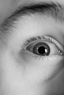
Human eye, infrared image C013 / 7322
MODEL RELEASED. Human eye, close-up infrared image. This is the right-hand eye of a 21-year-old man, with raised eyebrow and distended pupil

Background imageBlood Vessels Collection: Human arm, infrared image C013 / 7325

Human arm, infrared image C013 / 7325
MODEL RELEASED. Human arm. Infrared image of the anterior surface of the right-hand arm of a 24-year-old man. Infrared lighting can be used to visualize subcutaneous structures

Background imageBlood Vessels Collection: Cast of a human brain C013 / 6545

Cast of a human brain C013 / 6545
Cast of a human brain

Background imageBlood Vessels Collection: Arm anatomy, artwork C013 / 4582

Arm anatomy, artwork C013 / 4582
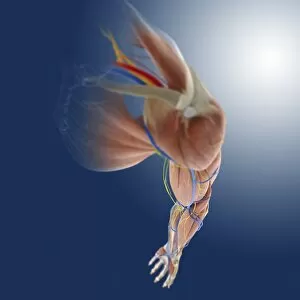
Arm anatomy. Computer artwork of an oblique anterior view looking down a human arm, showing muscles and tendons (brown and white), bones and ligaments (white), arteries and veins (red and blue)

Background imageBlood Vessels Collection: Leg anatomy, artwork C013 / 4497

Leg anatomy, artwork C013 / 4497
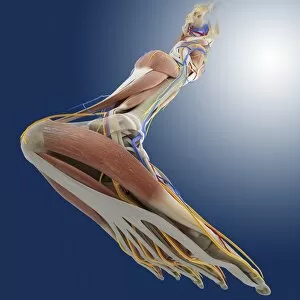
Leg anatomy. Computer artwork of an oblique posterior view looking up a human leg, showing muscles and tendons (brown and white), bones and ligaments (white), arteries and veins (red and blue)

Background imageBlood Vessels Collection: Leg anatomy, artwork C013 / 4495

Leg anatomy, artwork C013 / 4495
Leg anatomy. Computer artwork of an oblique side view looking down a human leg, showing muscles and tendons (brown and white), bones and ligaments (white), arteries and veins (red and blue)

Background imageBlood Vessels Collection: Leg anatomy, artwork C013 / 4496

Leg anatomy, artwork C013 / 4496
Leg anatomy. Computer artwork of an oblique anterior view looking up a human leg, showing muscles and tendons (brown and white), bones and ligaments (white), arteries and veins (red and blue)

Background imageBlood Vessels Collection: Leg anatomy, artwork C013 / 4493

Leg anatomy, artwork C013 / 4493
Leg anatomy. Computer artwork of an oblique anterior view looking down a human leg, showing muscles and tendons (brown and white), bones and ligaments (white), arteries and veins (red and blue)

Background imageBlood Vessels Collection: Leg anatomy, artwork C013 / 4494

Leg anatomy, artwork C013 / 4494
Leg anatomy. Computer artwork of an oblique anterior view looking down a human leg, showing muscles and tendons (brown and white), bones and ligaments (white), arteries and veins (red and blue)

Background imageBlood Vessels Collection: Leg ligaments, artwork

Leg ligaments, artwork
Leg anatomy. Computer artwork of an anterior view of a human leg, showing the muscles and tendons (brown and white), bones and ligaments (white), arteries and veins (red and blue), nerves (yellow)

Background imageBlood Vessels Collection: Cardiovascular system, artwork C013 / 4413

Cardiovascular system, artwork C013 / 4413
Cardiovascular system, computer artwork. The heart (centre, sectioned) is a hollow muscle that pumps blood around the body. Arteries, which carry oxygenated blood around the body, are red

Background imageBlood Vessels Collection: Heart-lung system, artwork C013 / 4412

Heart-lung system, artwork C013 / 4412
Heart-lung system, computer artwork. The heart (centre) is a hollow muscle that pumps blood around the body. Arteries, which carry oxygenated blood around the body, are red

Background imageBlood Vessels Collection: Heart-lung system, artwork C013 / 4411

Heart-lung system, artwork C013 / 4411
Heart-lung system, computer artwork. The heart (centre) is a hollow muscle that pumps blood around the body. Arteries, which carry oxygenated blood around the body, are red

Background imageBlood Vessels Collection: Heart-lung system, artwork C013 / 4410

Heart-lung system, artwork C013 / 4410
Heart-lung system, computer artwork. The heart (lower centre) is a hollow muscle that pumps blood around the body. Arteries, which carry oxygenated blood around the body, are red

Background imageBlood Vessels Collection: Heart-lung system, artwork C013 / 4408

Heart-lung system, artwork C013 / 4408
Heart-lung system, computer artwork. The heart (centre) is a hollow muscle that pumps blood around the body. Arteries, which carry oxygenated blood around the body, are red

Background imageBlood Vessels Collection: Heart-lung system, artwork C013 / 4409

Heart-lung system, artwork C013 / 4409
Heart-lung system, computer artwork. The heart (centre left) is a hollow muscle that pumps blood around the body. Arteries, which carry oxygenated blood around the body, are red

Background imageBlood Vessels Collection: Heart-lung system, artwork C013 / 4407

Heart-lung system, artwork C013 / 4407
Heart-lung system, computer artwork. The heart (centre left) is a hollow muscle that pumps blood around the body. Arteries, which carry oxygenated blood around the body, are red

Background imageBlood Vessels Collection: Cardiovascular system, artwork C013 / 4406

Cardiovascular system, artwork C013 / 4406
Cardiovascular system, computer artwork. The heart (centre) is a hollow muscle that pumps blood around the body. Arteries, which carry oxygenated blood around the body, are red

Background imageBlood Vessels Collection: Cardiovascular system, artwork C013 / 4405

Cardiovascular system, artwork C013 / 4405
Cardiovascular system, computer artwork. The heart (centre) is a hollow muscle that pumps blood around the body. Arteries, which carry oxygenated blood around the body, are red

Background imageBlood Vessels Collection: Heart and coronary arteries, artwork C013 / 4718

Heart and coronary arteries, artwork C013 / 4718
Heart and coronary arteries. 3D computer artwork of the external anatomy of a human heart. The surface blood vessels are the coronary arteries (red) and veins (pink)

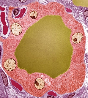
Background imageBlood Vessels Collection: Thyroid gland follicle, TEM

Thyroid gland follicle, TEM
Thyroid gland follicle. Coloured transmission electron micrograph (TEM) of a follicle from the thyroid gland. The follicle consists of a layer of epithelial cells (pink)

Background imageBlood Vessels Collection: Male anatomy

Male anatomy
MODEL RELEASED. Male anatomy. Man overlaid with an artwork of his internal organs and blood vessels. The heart is seen in the centre of his upper chest

Background imageBlood Vessels Collection: Upper body anatomy

Upper body anatomy
MODEL RELEASED. Upper body anatomy. Mans upper body overlaid with an artwork of the heart and the blood vessels and musculature of the chest and neck

Background imageBlood Vessels Collection: Kidneys and adrenal glands

Kidneys and adrenal glands
Kidneys. Computer artwork of the human renal system. The kidneys (brown/red, left and right) filter waste products and water from blood that passes through the organs in the renal arteries (red)

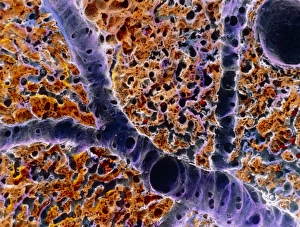
Background imageBlood Vessels Collection: False-colour SEM of vessels in liver lobule

False-colour SEM of vessels in liver lobule
Liver tissue. False-colour scanning electron micrograph (SEM) of blood vessels around a lobule, the functional unit of the liver

Background imageBlood Vessels Collection: Renal blood vessels, X-ray

Renal blood vessels, X-ray
Renal blood vessels. Angiogram (X-ray) of blood vessels in a healthy kidney. The large artery entering the kidney is the renal artery, which branches into many smaller capillaries inside the kidney

Background imageBlood Vessels Collection: Pancreas

Pancreas. Computer artwork of a human pancreas. The pancreas is between 15 and 20 centimetres in length. It is located in the abdominal cavity behind the stomach

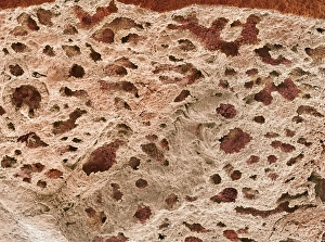
Background imageBlood Vessels Collection: Lung alveoli, SEM

Lung alveoli, SEM
Lung alveoli. Coloured scanning electron micrograph (SEM) of alveoli (air spaces) in the lungs. Blood vessels containing erythrocytes (red blood cells) are also seen

Background imageBlood Vessels Collection: Kidney

Kidney

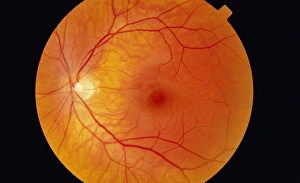
Background imageBlood Vessels Collection: Healthy retina

Healthy retina
Normal retina, ophthalmoscope image. The retina is the light-sensitive membrane that lines the back of the eye. Blood vessels (red) radiate out from the optic disc (yellow, left)

Background imageBlood Vessels Collection: Human blood vessels

Human blood vessels. Historical artwork of the blood vessels in a human body. The outlines of the arms and legs are clearly seen, and the main body artery (the aorta) runs down centre

Background imageBlood Vessels Collection: Skin anatomy

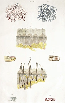
Skin anatomy. Historical anatomical artwork of the human skin. The main diagram (lower centre) shows scalp hairs and sebaceous glands (yellow, near surface)

Background imageBlood Vessels Collection: Female pelvis, coloured 3-D CT scan

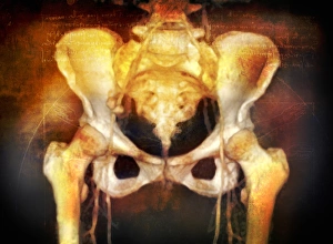
Female pelvis, coloured 3-D CT scan
Female pelvis, coloured 3-D computed tomography (CT) scan. The upper part of each thigh bone (femur) is seen at each side of the pelvis, where they form the hip joints

Background imageBlood Vessels Collection: Torso blood vessels

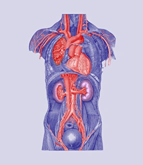
Torso blood vessels. Historical artwork of a human torso that has been dissected to show major blood vessels. The heart (upper centre) pumps blood to the lungs on either side (to be oxygenated)

Background imageBlood Vessels Collection: Coloured SEM of blood vessels in the skin

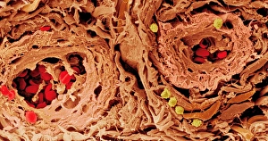
Coloured SEM of blood vessels in the skin
Skin blood vessels. Coloured scanning electron micrograph (SEM) of two small blood vessels (far left and upper right) in the dermis of the skin


All Professionally Made to Order for Quick Shipping

Blood vessels are the intricate network of highways that carry life-sustaining blood throughout our bodies. From the brain to the heart, from the retina to the neck and shoulder arteries, they ensure that every organ receives vital nutrients and oxygen. The 3D angiogram C007 / 1981 reveals a mesmerizing view of brain blood vessels, showcasing their complexity and importance. The heart, often referred to as the powerhouse of our body, relies on a vast system to pump oxygenated blood to every corner. Meanwhile, X-ray images capture the intricacies of neck and shoulder arteries, reminding us how crucial these pathways are for maintaining overall health. Intriguingly detailed artwork showcases both brain anatomy and male groin arteries from an 1825 perspective. These depictions serve as a testament to humanity's long-standing fascination with understanding our own bodies. Anatomical artwork C013 / 7419 beautifully illustrates arm circulation while highlighting its significance in ensuring proper functioning. Similarly captivating is pancreas anatomy depicted in yet another stunning piece of artistry. Head and chest arteries portrayed in an 1825 artwork remind us that even centuries ago, humans recognized the importance of studying these essential conduits for sustaining life. Migraine pain serves as a stark reminder that when something goes awry within our intricate vascular system, they can have debilitating consequences. These glimpses into human anatomy showcase not only scientific knowledge but also artistic expression at its finest. Finally, abdominal arteries captured through X-ray P206 / 0309 provide insight into yet another complex region where blood vessels play a critical role in maintaining optimal health. Blood vessels truly embody the essence of human existence - connecting each part like threads woven together in an intricate tapestry. Understanding their structure and function allows us to appreciate just how remarkable our bodies are; delicate yet resilient systems working tirelessly behind-the-scenes to keep us alive and thriving.