
Blood Vessels Collection (page 5)
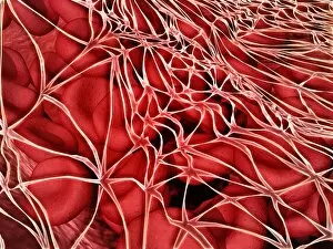
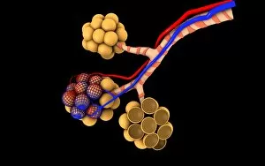
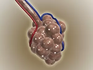
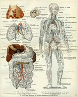
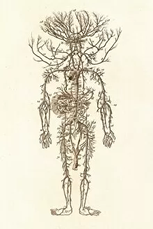
Blood vessels are the intricate network of highways that carry life-sustaining blood throughout our bodies


All Professionally Made to Order for Quick Shipping
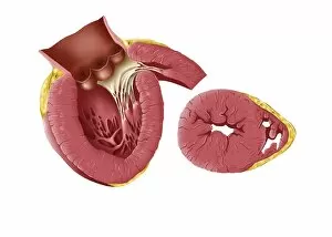
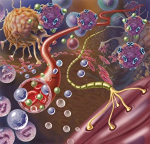
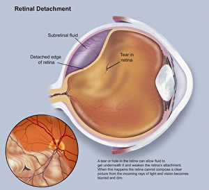
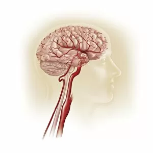
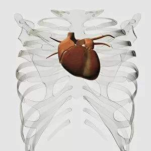
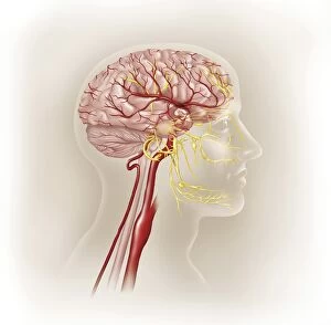
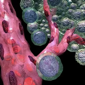
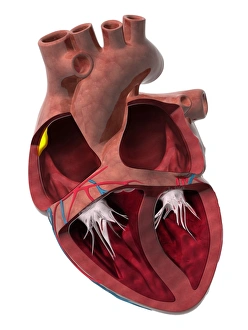
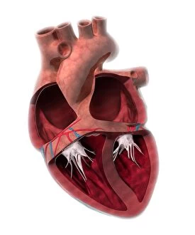
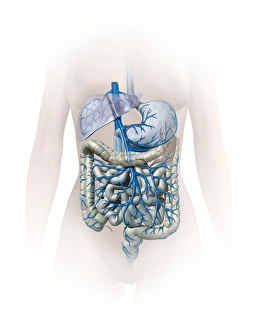
Blood vessels are the intricate network of highways that carry life-sustaining blood throughout our bodies. From the brain to the heart, from the retina to the neck and shoulder arteries, they ensure that every organ receives vital nutrients and oxygen. The 3D angiogram C007 / 1981 reveals a mesmerizing view of brain blood vessels, showcasing their complexity and importance. The heart, often referred to as the powerhouse of our body, relies on a vast system to pump oxygenated blood to every corner. Meanwhile, X-ray images capture the intricacies of neck and shoulder arteries, reminding us how crucial these pathways are for maintaining overall health. Intriguingly detailed artwork showcases both brain anatomy and male groin arteries from an 1825 perspective. These depictions serve as a testament to humanity's long-standing fascination with understanding our own bodies. Anatomical artwork C013 / 7419 beautifully illustrates arm circulation while highlighting its significance in ensuring proper functioning. Similarly captivating is pancreas anatomy depicted in yet another stunning piece of artistry. Head and chest arteries portrayed in an 1825 artwork remind us that even centuries ago, humans recognized the importance of studying these essential conduits for sustaining life. Migraine pain serves as a stark reminder that when something goes awry within our intricate vascular system, they can have debilitating consequences. These glimpses into human anatomy showcase not only scientific knowledge but also artistic expression at its finest. Finally, abdominal arteries captured through X-ray P206 / 0309 provide insight into yet another complex region where blood vessels play a critical role in maintaining optimal health. Blood vessels truly embody the essence of human existence - connecting each part like threads woven together in an intricate tapestry. Understanding their structure and function allows us to appreciate just how remarkable our bodies are; delicate yet resilient systems working tirelessly behind-the-scenes to keep us alive and thriving.