
Blood Sucker Collection
"Blood Suckers: Unveiling the Tiny Terrors of Nature" In this captivating journey through the microscopic world


All Professionally Made to Order for Quick Shipping
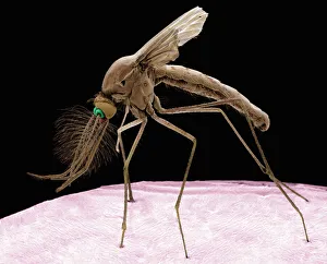
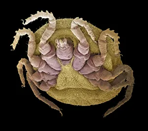
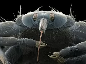
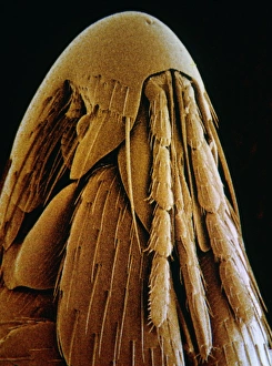
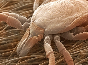
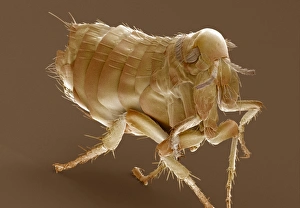
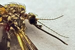
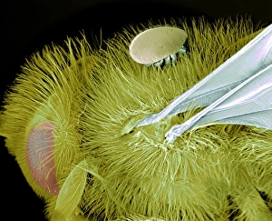
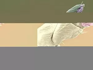
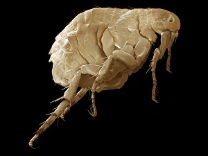
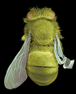
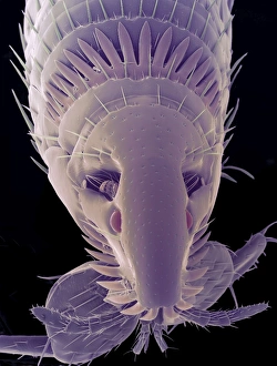
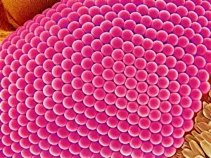
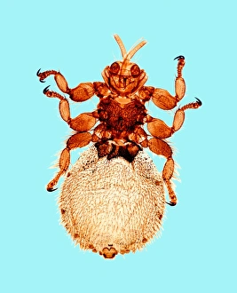
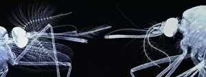
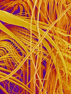
"Blood Suckers: Unveiling the Tiny Terrors of Nature" In this captivating journey through the microscopic world, we encounter some notorious blood-sucking creatures that have both fascinated and plagued humanity for centuries. First up is the Culex mosquito, a common culprit behind those irritating itchy bites. Underneath an electron microscope (SEM), its delicate body reveals intricate details, reminding us of its stealthy nature. Moving on to the sheep tick, another formidable bloodsucker lurking in grassy fields. Its SEM image showcases its claw-like appendages designed to latch onto unsuspecting hosts with precision and efficiency. Amidst these tiny terrors lies breathtaking scenery that contrasts their sinister reputation – a vibrant color lithograph captures nature's beauty at its finest. It serves as a reminder that even amidst danger, there is always something awe-inspiring to behold. However, not everything is as it seems. The liar among them emerges – a female mosquito head under SEM reveals her deceptive tactics used to extract precious blood while remaining undetected by our senses. Enter the black horse fly C018/4697; with its menacing appearance and sharp mouthparts ready for action, it leaves no doubt about its role as one of nature's most fearsome bloodsuckers. Transitioning from insects to fleas, we explore their microscopic world through light microscopy (LM). A detailed LM image of bird flea showcases their ability to adapt perfectly to avian hosts while causing discomfort and irritation when they occasionally cross paths with humans. Zooming in further under SEM lenses unveils intricate structures on the bird flea's head - an astonishing display of evolution's craftsmanship tailored specifically for extracting nourishment from feathered creatures. Returning back to terrestrial parasites, we revisit the sheep tick under SEM once more. Its resilient exoskeleton reminds us how these tiny arachnids can survive harsh conditions while posing health risks for both animals and humans.