Fraud Blocker Skip to main content

Blood Flow Collection (page 4)

"Unveiling the Intricate Pathways: Exploring Blood Flow Through Time" Step back in time to 1861 with this captivating hand-coloured engraving

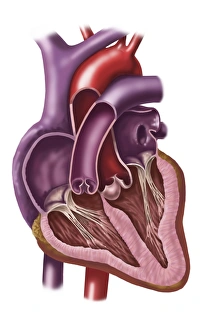
Background imageBlood Flow Collection: Interior of human heart showing atria and ventricles

Interior of human heart showing atria and ventricles

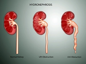
Background imageBlood Flow Collection: Hydronephrosis condition of the kidney

Hydronephrosis condition of the kidney
Hydronephrosis is a condition that typically occurs when one kidney becomes swollen due to the failure of normal drainage of urine from the kidney to the bladder

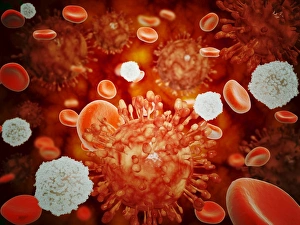
Background imageBlood Flow Collection: Microscopic view of H5N1 virus with red blood cells and white blood cells

Microscopic view of H5N1 virus with red blood cells and white blood cells

Background imageBlood Flow Collection: Conceptual image of chromosomes inside the blood stream

Conceptual image of chromosomes inside the blood stream

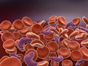
Background imageBlood Flow Collection: Conceptual image of sickle cell anemia with red blood cells

Conceptual image of sickle cell anemia with red blood cells

Background imageBlood Flow Collection: Conceptual image of a red blood cell

Conceptual image of a red blood cell

Background imageBlood Flow Collection: Human kidney with artery

Human kidney with artery

Background imageBlood Flow Collection: Normal artery versus artery in spasm

Normal artery versus artery in spasm

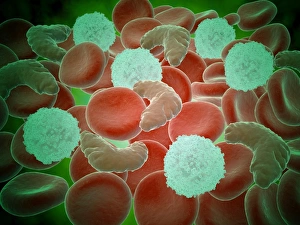
Background imageBlood Flow Collection: Sickle cell anemia with red blood cells and white bood cells

Sickle cell anemia with red blood cells and white bood cells
Conceptual image of sickle cell anemia with red blood cells and white bood cells

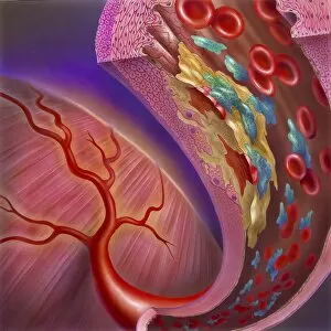
Background imageBlood Flow Collection: Artery showing atherosclerotic plaque, platelets and red blood cells

Artery showing atherosclerotic plaque, platelets and red blood cells
Close-up of artery showing atherosclerotic plaque, platelets and red blood cells

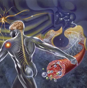
Background imageBlood Flow Collection: Schematic of the hypothalamus receiving nerve impulses from the body

Schematic of the hypothalamus receiving nerve impulses from the body and sending messages to the circulatory and nervous system

Background imageBlood Flow Collection: Arteriole with red blood cells, white blood cells and oxygen

Arteriole with red blood cells, white blood cells and oxygen

Background imageBlood Flow Collection: Microscopic view of fat plaque inside the artery

Microscopic view of fat plaque inside the artery

Background imageBlood Flow Collection: Microscopic view of blood flow inside an artery with stent deployment

Microscopic view of blood flow inside an artery with stent deployment

Background imageBlood Flow Collection: Female body sitting in dynamic posture with circulatory system superimposed

Female body sitting in dynamic posture with circulatory system superimposed
Human female body sitting in dynamic posture, with full circulatory system superimposed

Background imageBlood Flow Collection: Interior detail of human kidney

Interior detail of human kidney

Background imageBlood Flow Collection: Insertion of stent into atherosclerotic artery

Insertion of stent into atherosclerotic artery

Background imageBlood Flow Collection: Artery cross-section with blood flow, fat plaque and stent deployment

Artery cross-section with blood flow, fat plaque and stent deployment
Microscopic view of an artery cross-section with blood flow, fat plaque and stent deployment

Background imageBlood Flow Collection: Conceptual image of red blood cells with platelets

Conceptual image of red blood cells with platelets

Background imageBlood Flow Collection: Microscopic view of HIV virus inside the lungs

Microscopic view of HIV virus inside the lungs

Background imageBlood Flow Collection: Microscopic view of plasma cell inside blood vessel

Microscopic view of plasma cell inside blood vessel

Background imageBlood Flow Collection: Inside view of the artery with red blood cells

Inside view of the artery with red blood cells

Background imageBlood Flow Collection: Blood vessel anatomy

Blood vessel anatomy

Background imageBlood Flow Collection: Anatomy of human veins and arteries, black background

Anatomy of human veins and arteries, black background

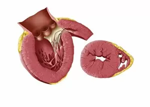
Background imageBlood Flow Collection: Enlarged left ventricle of the human heart

Enlarged left ventricle of the human heart

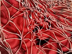
Background imageBlood Flow Collection: Conceptual image of red blood cells with fibrin

Conceptual image of red blood cells with fibrin

Background imageBlood Flow Collection: Conceptual image of sickle cell anemia

Conceptual image of sickle cell anemia. Sickle cell anemia is a disease in which your body produces abnormally shaped red blood cells. The cells are shaped like a crescent or sickle
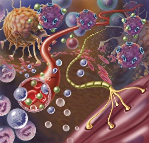
Background imageBlood Flow Collection: Nerve with myelin sheath, seen in lower right, connects with muscle

Nerve with myelin sheath, seen in lower right, connects with muscle. Blood vessel and immune cells are seen in the center and upper right of image

Background imageBlood Flow Collection: Urine production in human kidney

Urine production in human kidney

Background imageBlood Flow Collection: Human kidney

Human kidney

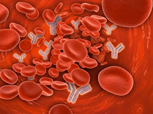
Background imageBlood Flow Collection: Microscopic view of human anitbodies with red blood cells

Microscopic view of human anitbodies with red blood cells. Human antibodies are the Y-shaped proteins used by the immune system to defend against foreign objects like bacteria and viruses

Background imageBlood Flow Collection: Three dimensional view of human heart, left side

Three dimensional view of human heart, left side

Background imageBlood Flow Collection: Microscopic view of sicke cells causing anemia disease

Microscopic view of sicke cells causing anemia disease

Background imageBlood Flow Collection: Cross-section of human kidney

Cross-section of human kidney

Background imageBlood Flow Collection: Microscopic view of blood cells

Microscopic view of blood cells

Background imageBlood Flow Collection: Conceptual image of a blood vessel

Conceptual image of a blood vessel

Background imageBlood Flow Collection: Conceptual image of red blood cells

Conceptual image of red blood cells

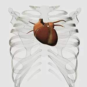
Background imageBlood Flow Collection: Medical illustration of human heart and rib cage

Medical illustration of human heart and rib cage, three dimensional view

Background imageBlood Flow Collection: Polycystic kidney

Polycystic kidney

Background imageBlood Flow Collection: Components of blood vessel interact with intracellular fluid

Components of blood vessel interact with intracellular fluid; white blood cells, red blood cells, oxygen, platelets and hormones

Background imageBlood Flow Collection: Ureterovesical junction (UVJ) in the kidney

Ureterovesical junction (UVJ) in the kidney. This is the point where the ureters from the kidney connect to the bladder

Background imageBlood Flow Collection: Malria spores in the human blood stream

Malria spores in the human blood stream
The malaria spores move from the mosquito to the human blood stream. the spores divide many times and produce large numbers of spores

Background imageBlood Flow Collection: Microscopic view of red blood cells flowing inside lungs

Microscopic view of red blood cells flowing inside lungs

Background imageBlood Flow Collection: Artery cross section with red blood cell flow

Artery cross section with red blood cell flow

Background imageBlood Flow Collection: Medical illustration of arteries, veins and lymphatic system in female chest area

Medical illustration of arteries, veins and lymphatic system in female chest area
Medical illustration of arteries, veins and lymphatic system with heart. Profile view of female chest area

Background imageBlood Flow Collection: Illustration of human heart

Illustration of human heart

Background imageBlood Flow Collection: Cross section of human respiratory system

Cross section of human respiratory system

Background imageBlood Flow Collection: Human kidney and adrenal gland

Human kidney and adrenal gland


All Professionally Made to Order for Quick Shipping

"Unveiling the Intricate Pathways: Exploring Blood Flow Through Time" Step back in time to 1861 with this captivating hand-coloured engraving, showcasing the marvels of the human circulatory system. Delve into a world where diagrams come alive, revealing the secrets of blood supply within our lungs. Embark on a journey through anatomy as you explore the intricate details of our feet - skin, veins, arteries, muscles and bones all working harmoniously to facilitate blood flow. Marvel at the complexity of our arm and hand's inner workings; behold an enlightening diagram displaying bones intertwined with veins and arteries. Witness firsthand how life thrives within us as you peer into a vein's depths - red and white blood cells dancing alongside platelets. Take a cross-sectional glimpse into one of nature's most vital organs –the Human Heart– discovering its awe-inspiring structure that keeps us alive. Prepare for an educational feast as we unravel every nook and cranny within this incredible organ. With labelled structures guiding your exploration, discover each component responsible for maintaining our heartbeat rhythmically. But let us not forget about other essential parts that contribute to our well-being. Dive deep into body anatomy featuring kidneys – these unsung heroes tirelessly filtering waste from our bloodstream. Marvel at female physiology as we reveal both digestive and circulatory systems intertwining seamlessly beneath her skin. Witness internal organs coming together in perfect harmony –a testament to nature's remarkable design. Finally, immerse yourself in heart interior like never before with a frontal section view. Uncover its hidden chambers and valves intricately arranged to ensure efficient blood circulation throughout our bodies. Join us on this extraordinary voyage through time as we celebrate the wonders captured in stunning illustrations from centuries past. Prepare to be amazed by the gross anatomy that sustains human life itself.