
Blood Flow Collection (page 3)
"Unveiling the Intricate Pathways: Exploring Blood Flow Through Time" Step back in time to 1861 with this captivating hand-coloured engraving


All Professionally Made to Order for Quick Shipping
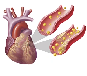
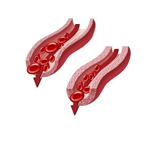
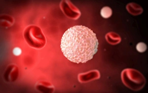
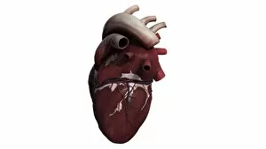
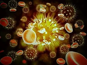
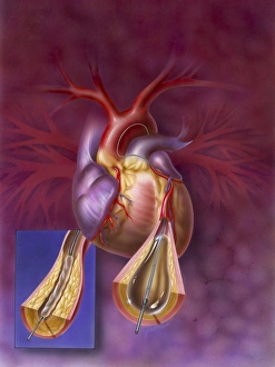
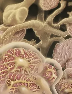
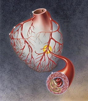
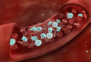
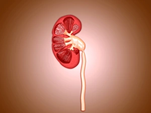
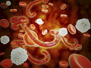
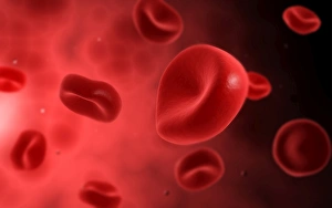
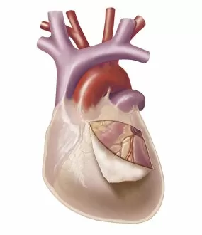
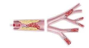
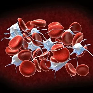
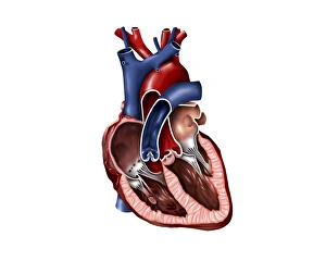
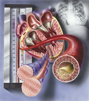
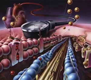
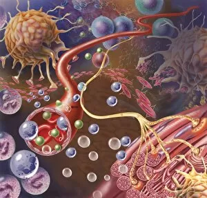
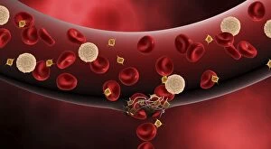
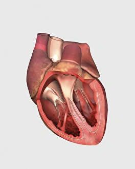
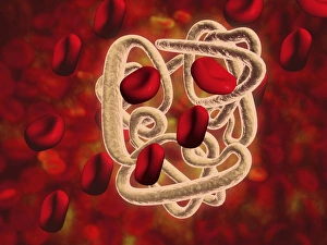
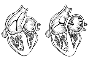
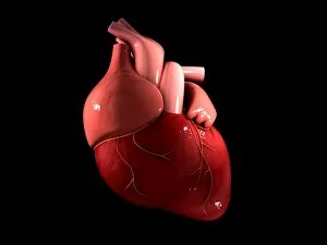
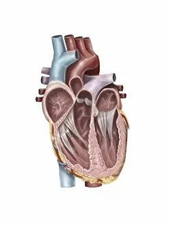
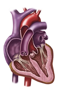
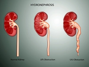
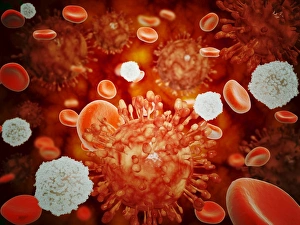
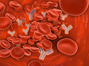
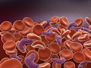
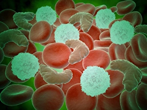
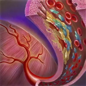
"Unveiling the Intricate Pathways: Exploring Blood Flow Through Time" Step back in time to 1861 with this captivating hand-coloured engraving, showcasing the marvels of the human circulatory system. Delve into a world where diagrams come alive, revealing the secrets of blood supply within our lungs. Embark on a journey through anatomy as you explore the intricate details of our feet - skin, veins, arteries, muscles and bones all working harmoniously to facilitate blood flow. Marvel at the complexity of our arm and hand's inner workings; behold an enlightening diagram displaying bones intertwined with veins and arteries. Witness firsthand how life thrives within us as you peer into a vein's depths - red and white blood cells dancing alongside platelets. Take a cross-sectional glimpse into one of nature's most vital organs –the Human Heart– discovering its awe-inspiring structure that keeps us alive. Prepare for an educational feast as we unravel every nook and cranny within this incredible organ. With labelled structures guiding your exploration, discover each component responsible for maintaining our heartbeat rhythmically. But let us not forget about other essential parts that contribute to our well-being. Dive deep into body anatomy featuring kidneys – these unsung heroes tirelessly filtering waste from our bloodstream. Marvel at female physiology as we reveal both digestive and circulatory systems intertwining seamlessly beneath her skin. Witness internal organs coming together in perfect harmony –a testament to nature's remarkable design. Finally, immerse yourself in heart interior like never before with a frontal section view. Uncover its hidden chambers and valves intricately arranged to ensure efficient blood circulation throughout our bodies. Join us on this extraordinary voyage through time as we celebrate the wonders captured in stunning illustrations from centuries past. Prepare to be amazed by the gross anatomy that sustains human life itself.