Fraud Blocker Skip to main content

Bacterial Collection (page 9)

"Bacterial: Unveiling the Microscopic World of Disease and Discovery" Step into a time capsule as we journey through history, exploring the captivating realm of bacteria

Background imageBacterial Collection: Bacterial outer membrane protein molecule C014 / 4949

Bacterial outer membrane protein molecule C014 / 4949
Bacterial outer membrane protein molecule. Computer model showing the secondary structure of a molecule of outer membrane transporter FecA protein from Escherichia coli (E. coli) bacteria

Background imageBacterial Collection: Hand bacteria C014 / 4839

Hand bacteria C014 / 4839
Hand bacteria. Colony of bacteria that have grown where a hand was pressed onto an agar plate

Background imageBacterial Collection: Anthrax lethal factor molecule

Anthrax lethal factor molecule
Anthrax lethal factor, molecular model. This enzyme is one of three protein components that form the anthrax toxin produced by the bacterium Bacillus anthracis

Background imageBacterial Collection: Robert Cooley, US entomologist

Robert Cooley, US entomologist
Robert Allen Cooley (1873-1968), US entomologist, in his laboratory. Cooley was professor of zoology and entomology at Montana State University, USA

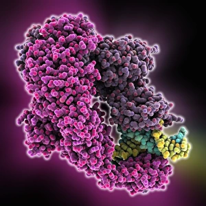
Background imageBacterial Collection: E. coli DNA mismatch repair complex

E. coli DNA mismatch repair complex. Computer model showing the structure of a MutS (Mutator S) protein from Escherichia coli (E)

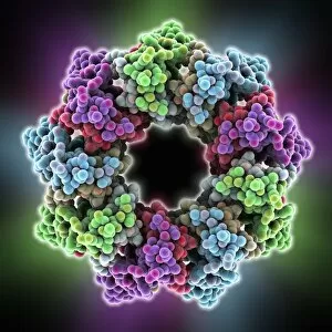
Background imageBacterial Collection: Light-harvesting protein complex

Light-harvesting protein complex, molecular model. Peripheral light-harvesting protein complex from the purple bacterium Rhodopseudomonas acidophila

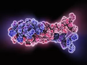
Background imageBacterial Collection: Bacterial RNA-binding protein molecule

Bacterial RNA-binding protein molecule
Bacterial RNA-binding protein. Molecular model of the bacterial RNA-binding protein Hfq complexed with RNA (ribonucleic acid)

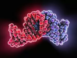
Background imageBacterial Collection: Bacterial RNA plasmid loop-loop complex

Bacterial RNA plasmid loop-loop complex, molecular model. This strand of ribonucleic acid (RNA) is part of a plasmid, the loop of genetic material found in bacterial cells

Background imageBacterial Collection: Methane monooxygenase enzyme

Methane monooxygenase enzyme, molecular model. This is the particulate methane monooxygenase (pMMO) form of this metalloenzyme, an integral membrane protein that contains copper and zinc

Background imageBacterial Collection: Shark-skin-inspired antibacterial surface

Shark-skin-inspired antibacterial surface. Close-up of Sharklet an antibacterial material whose surface structure was inspired by the microstructures found on the surface of shark skin

Background imageBacterial Collection: H-NS chromatin-structuring protein

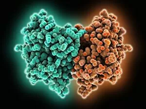
H-NS chromatin-structuring protein. Molecular model of the oligomerization domain of the H-NS protein from the Escherichia coli bacterium. This dimeric molecule folds in on itself, as shown here

Background imageBacterial Collection: Bacterial regulator-DNA complex

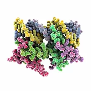
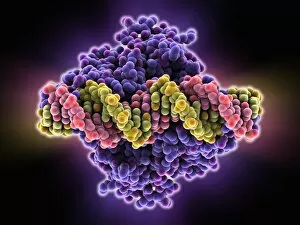
Bacterial regulator-DNA complex. Molecular model of a complex formed between a bacterial regulator called SarA (orange and brown) and a fragment of DNA (pink and yellow-green strands)

Background imageBacterial Collection: Bacterial protein-chaperone complex

Bacterial protein-chaperone complex. Molecular model of a bacterial effector protein binding to a chaperone protein that helps prevent keep the bacterial protein in an unfolded or partially folded

Background imageBacterial Collection: Restriction enzyme cutting DNA

Restriction enzyme cutting DNA
Fragment of DNA bound by the restriction endonucleaseEcoRI. The protein is a dimer, with each subunitable to bind and cut one strand of DNA

Background imageBacterial Collection: Bacterial transcription factor molecule C016 / 2061

Bacterial transcription factor molecule C016 / 2061
Bacterial transcription factor. Molecular model of the transcription factor AmrZ from the bacterium Pseudomonas aeruginosa complexed with DNA (deoxyribonucleic acid)

Background imageBacterial Collection: Ribosomal RNA-binding protein molecule

Ribosomal RNA-binding protein molecule. Computer model showing the structure of a ribosomal protein L9 (RPL9) molecule from Bacillus stearothermophilus bacteria

Background imageBacterial Collection: Lumazine synthase molecule

Lumazine synthase molecule. Computer model showing the structure of a lumazine synthase enzyme molecule from a Brucella abortus bacterium

Background imageBacterial Collection: Bacterial alpha-hemolysin toxin C015 / 7067

Bacterial alpha-hemolysin toxin C015 / 7067
Bacterial alpha-hemolysin toxin, molecular model. This toxin is secreted by the bacterium Staphylococcus aureus. It is an example of a pore-forming toxin

Background imageBacterial Collection: Mandelate racemase enzyme C015 / 7021

Mandelate racemase enzyme C015 / 7021
Mandelate racemase enzyme, molecular model. This bacterial enzyme is an example of a muconate lactonizing enzyme, and plays a key role in the metabolism of soil and aquatic bacteria

Background imageBacterial Collection: Mandelate racemase enzyme C015 / 7020

Mandelate racemase enzyme C015 / 7020
Mandelate racemase enzyme, molecular model. This bacterial enzyme is an example of a muconate lactonizing enzyme, and plays a key role in the metabolism of soil and aquatic bacteria

Background imageBacterial Collection: Historical iron corrosion samples C016 / 2040

Historical iron corrosion samples C016 / 2040
Historical iron corrosion samples. Series of glass tubes with 70-year-old samples of iron under different conditions, demonstrating the effect on corrosion. The years vary between 1907 and 1910

Background imageBacterial Collection: Restriction enzyme and DNA C015 / 6941

Restriction enzyme and DNA C015 / 6941
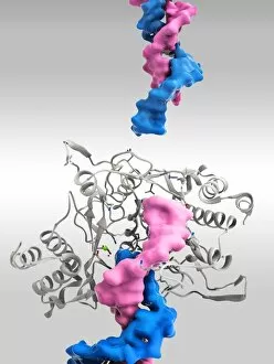
Restriction enzyme and DNA. Molecular model showing an EcoRI endonuclease enzyme (purple) bound to a DNA (deoxyribonucleic acid) strand (blue). EcoRI is an enzyme isolated from strains of E

Background imageBacterial Collection: NpmA methyltransferase C016 / 2031

NpmA methyltransferase C016 / 2031
NpmA methyltransferase, molecular model. Methyltransferase enzymes act to add methyl groups to nucleic acids such as DNA, a process called DNA methylation

Background imageBacterial Collection: NpmA methyltransferase C016 / 2030

NpmA methyltransferase C016 / 2030
NpmA methyltransferase, molecular model. Methyltransferase enzymes act to add methyl groups to nucleic acids such as DNA, a process called DNA methylation

Background imageBacterial Collection: Pond bacteria C015 / 6371

Pond bacteria C015 / 6371
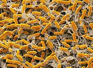
Pond bacteria. Coloured scanning electron micrograph (SEM) of rod-shaped bacteria (bacilli) from a freshwater pond. Magnification: x4000, when printed 10cm wide

Background imageBacterial Collection: Pond bacteria C015 / 6369

Pond bacteria C015 / 6369
Pond bacteria. Coloured scanning electron micrograph (SEM) of rod-shaped bacteria (bacilli) from a freshwater pond. Magnification: x4000, when printed 10cm wide

Background imageBacterial Collection: C3-degrading proteinase enzyme C016 / 1363

C3-degrading proteinase enzyme C016 / 1363
C3-degrading proteinase enzyme. Molecular model of a proteinase enzyme from the TIGR4 strain of the bacterium Streptococcus pneumoniae

Background imageBacterial Collection: C3-degrading proteinase enzyme C016 / 1362

C3-degrading proteinase enzyme C016 / 1362
C3-degrading proteinase enzyme. Molecular model of a proteinase enzyme from the TIGR4 strain of the bacterium Streptococcus pneumoniae

Background imageBacterial Collection: Acyl-CoA hydrolase enzyme C015 / 6724

Acyl-CoA hydrolase enzyme C015 / 6724
Acyl-CoA hydrolase enzyme, molecular model. This is an acyl-CoA thioesterase enzyme. Thioesterases hydrolyze coenzyme A (CoA) esters, producing the free acid and CoA

Background imageBacterial Collection: Acyl-CoA hydrolase enzyme C015 / 6725

Acyl-CoA hydrolase enzyme C015 / 6725
Acyl-CoA hydrolase enzyme, molecular model. This is an acyl-CoA thioesterase enzyme. Thioesterases hydrolyze coenzyme A (CoA) esters, producing the free acid and CoA

Background imageBacterial Collection: Macrophage engulfing TB bacteria, SEM C014 / 0596

Macrophage engulfing TB bacteria, SEM C014 / 0596
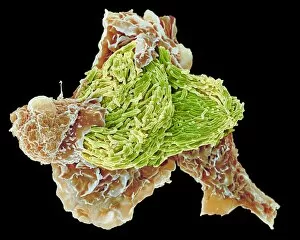
Macrophage engulfing TB bacteria. Coloured scanning electron micrograph (SEM) of a macrophage white blood cell engulfing a tuberculosis (Mycobacterium tuberculosis) bacterium (orange)

Background imageBacterial Collection: Macrophage engulfing TB bacteria, SEM C014 / 0598

Macrophage engulfing TB bacteria, SEM C014 / 0598
Macrophage engulfing TB bacteria. Coloured scanning electron micrograph (SEM) of a macrophage white blood cell engulfing a tuberculosis (Mycobacterium tuberculosis) bacterium (purple)

Background imageBacterial Collection: Macrophage engulfing TB bacteria, SEM C014 / 0595

Macrophage engulfing TB bacteria, SEM C014 / 0595
Macrophage engulfing TB bacteria. Coloured scanning electron micrograph (SEM) of a macrophage white blood cell engulfing a tuberculosis (Mycobacterium tuberculosis) bacterium (pink)

Background imageBacterial Collection: Macrophage engulfing TB bacteria, SEM C014 / 0594

Macrophage engulfing TB bacteria, SEM C014 / 0594
Macrophage engulfing TB bacteria. Coloured scanning electron micrograph (SEM) of a macrophage white blood cell engulfing a tuberculosis (Mycobacterium tuberculosis) bacterium (yellow)

Background imageBacterial Collection: Macrophage engulfing TB bacteria, SEM C014 / 0593

Macrophage engulfing TB bacteria, SEM C014 / 0593
Macrophage engulfing TB bacteria. Coloured scanning electron micrograph (SEM) of a macrophage white blood cell engulfing a tuberculosis (Mycobacterium tuberculosis) bacterium (purple)

Background imageBacterial Collection: Aquaporin membrane protein C015 / 5922

Aquaporin membrane protein C015 / 5922
Aquaporin membrane protein, molecular model. Aquaporins are membrane proteins that form channels (lower right) that help water molecules pass in and out of cells

Background imageBacterial Collection: Macrophage engulfing TB bacteria, SEM C014 / 0592

Macrophage engulfing TB bacteria, SEM C014 / 0592
Macrophage engulfing TB bacteria. Coloured scanning electron micrograph (SEM) of a macrophage white blood cell engulfing a tuberculosis (Mycobacterium tuberculosis) bacterium (orange)

Background imageBacterial Collection: Macrophage engulfing TB bacteria, SEM C014 / 0590

Macrophage engulfing TB bacteria, SEM C014 / 0590
Macrophage engulfing TB bacteria. Coloured scanning electron micrograph (SEM) of a macrophage white blood cell engulfing a tuberculosis (Mycobacterium tuberculosis) bacterium (blue)

Background imageBacterial Collection: Transcription repressor protein and DNA C015 / 5810

Transcription repressor protein and DNA C015 / 5810
Transcription repressor protein and DNA, molecular model. The repressor protein (green) is binding to a strand of DNA (deoxyribonucleic acid, pink and purple)

Background imageBacterial Collection: Macrophage engulfing TB bacteria, SEM C014 / 0589

Macrophage engulfing TB bacteria, SEM C014 / 0589
Macrophage engulfing TB bacteria. Coloured scanning electron micrograph (SEM) of a macrophage white blood cell engulfing a tuberculosis (Mycobacterium tuberculosis) bacterium (yellow)

Background imageBacterial Collection: Macrophage engulfing TB bacteria, SEM C014 / 0591

Macrophage engulfing TB bacteria, SEM C014 / 0591
Macrophage engulfing TB bacteria. Coloured scanning electron micrograph (SEM) of a macrophage white blood cell engulfing a tuberculosis (Mycobacterium tuberculosis) bacterium (bright green)

Background imageBacterial Collection: Vitamin B12 transport protein C015 / 5824

Vitamin B12 transport protein C015 / 5824
Vitamin B12 transport protein, molecular model. This transmembrane protein, known as BTUB, is from the Escherichia coli bacterium

Background imageBacterial Collection: Checking the safety of milk

Checking the safety of milk
Milk production. View of a petri dish in which a sample of milk has been incubated. This is to ensure that the milk is safe to process for human consumption

Background imageBacterial Collection: Vitamin B12 transport protein C015 / 5823

Vitamin B12 transport protein C015 / 5823
Vitamin B12 transport protein, molecular model. This transmembrane protein, known as BTUB, is from the Escherichia coli bacterium

Background imageBacterial Collection: Transcription repressor protein and DNA C015 / 5811

Transcription repressor protein and DNA C015 / 5811
Transcription repressor protein and DNA, molecular model. The repressor protein (purple) is binding to a strand of DNA (deoxyribonucleic acid, yellow and pink)

Background imageBacterial Collection: Outer membrane phospholipase A molecule C015 / 6111

Outer membrane phospholipase A molecule C015 / 6111
Outer membrane phospholipase A. Molecular model of the integral membrane protein, outer membrane phospholipase A from the Escherichia coli bacterium

Background imageBacterial Collection: Actin-based motility, SEM C017 / 8335

Actin-based motility, SEM C017 / 8335
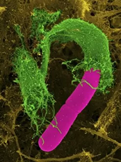
Actin-based motility. Coloured scanning electron micrograph (SEM) of Shigella sp. bacteria (pink) infecting cells. Filaments of the protein actin, which is part of the cells cystoskeleton

Background imageBacterial Collection: Actin-based motility, SEM C017 / 8334

Actin-based motility, SEM C017 / 8334
Actin-based motility. Coloured scanning electron micrograph (SEM) of Shigella sp. bacteria (pink) infecting cells. Filaments of the protein actin, which is part of the cells cystoskeleton


All Professionally Made to Order for Quick Shipping

"Bacterial: Unveiling the Microscopic World of Disease and Discovery" Step into a time capsule as we journey through history, exploring the captivating realm of bacteria. From the haunting presence of plague doctors in 17th-century artwork to cutting-edge scientific images captured by scanning electron microscopes (SEM), prepare to be fascinated by these tiny yet formidable organisms. Witness the battle between our immune system and Methicillin-resistant Staphylococcus aureus (MRSA) as a brave neutrophil engulfs it under SEM C018/8596. Marvel at the groundbreaking Calots spinal surgery from the 19th century, which revolutionized medical practices despite lurking bacterial threats. Peering through an X-ray, tuberculosis reveals its sinister grip on human lungs while skin disorders come alive in vivid artwork, reminding us of bacteria's impact on our bodies. E. coli bacteria, magnified under SEM, showcases their intricate structures that belie their harmful potential. Travel back to Soviet Russia in 1967 with a cholera vaccination poster that symbolizes humanity's fight against bacterial epidemics. Observe salmonella bacteria under SEM; their deceptively beautiful appearance belies their ability to cause severe illness. Delve into historical diagrams depicting anthrax cultures and learn about Yersinia pestis - responsible for devastating pandemics throughout history. The tuberculosis bacterium reminds us of its long-standing presence as one of humanity's greatest adversaries. Finally, witness nature's artistic flair with spiral spore chains formed by Streptomyces bacteria – showcasing both beauty and resilience within this microscopic world. Through these glimpses into bacterial realms past and present, we gain insight into our ongoing struggle against infectious diseases. Let this exploration ignite curiosity about microbial lifeforms that shape our existence – forever reminding us how knowledge can empower us in combating these invisible foes.