Fraud Blocker Skip to main content

Anatomy Collection (page 11)

"Unveiling the Intricacies of Anatomy: From Sensory Homunculus to Motor Homunculus" Step into the fascinating world of anatomy, where every detail tells a story

Background imageAnatomy Collection: Puer Cornutus : Puer cornutus: a child with horns. From the 1642 book Monstrorum Historia by

Puer Cornutus : Puer cornutus: a child with horns. From the 1642 book Monstrorum Historia by Ulisse Aldrovandi
UIG5327158 Puer Cornutus : Puer cornutus: a child with horns. From the 1642 book Monstrorum Historia by Ulisse Aldrovandi (Bologna, 1522-1605)

Background imageAnatomy Collection: Sus Capite & Pedibus Humanis : Sus capite & pedibus humanis, pig with human head and hands

Sus Capite & Pedibus Humanis : Sus capite & pedibus humanis, pig with human head and hands
UIG5327153 Sus Capite & Pedibus Humanis : Sus capite & pedibus humanis, pig with human head and hands. From the 1642 book Monstrorum Historia by Ulisse Aldrovandi (Bologna, 1522-1605)

Background imageAnatomy Collection: Pullus Equinus Humana Facie : Horse with human face. From the 1642 book Monstrorum Historia by

Pullus Equinus Humana Facie : Horse with human face. From the 1642 book Monstrorum Historia by Ulisse Aldrovandi
UIG5327149 Pullus Equinus Humana Facie : Horse with human face. From the 1642 book Monstrorum Historia by Ulisse Aldrovandi (Bologna, 1522-1605)

Background imageAnatomy Collection: Four Beasts : Hypertrichosis (also called Ambras syndrome) is an abnormal amount of hair growth

Four Beasts : Hypertrichosis (also called Ambras syndrome) is an abnormal amount of hair growth over the body;
UIG5327174 Four Beasts : Hypertrichosis (also called Ambras syndrome) is an abnormal amount of hair growth over the body; extensive cases of hypertrichosis have informally been called werewolf

Background imageAnatomy Collection: Agnus capite monstrifico : Agnus capite monstrifico, lamb with monstrous head

Agnus capite monstrifico : Agnus capite monstrifico, lamb with monstrous head
UIG5327169 Agnus capite monstrifico : Agnus capite monstrifico, lamb with monstrous head. From the 1642 book Monstrorum Historia by Ulisse Aldrovandi (Bologna, 1522-1605)

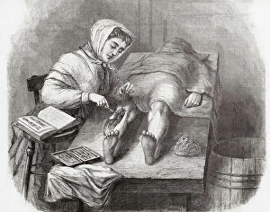
Background imageAnatomy Collection: Medical College for Women, New York

Medical College for Women, New York
7301631 Medical College for Women, New York.; Private Collection; (add.info.: A student dissecting a leg at the Medical College for Women on East Twelfth Street and Second Avenue, New York, USA)

Background imageAnatomy Collection: Anatomical section of the head

Anatomical section of the head
6266170 Anatomical section of the head; (add.info.: Anatomical section of the head. After a 19th century study by Robert Muys.); Photo © Ken Welsh. All rights reserved 2024.

Background imageAnatomy Collection: Lo spellato' (The bark) Bronze sculpture by Ludovico Cardi called Le Cigoli (1559-1613) Florence

Lo spellato' (The bark) Bronze sculpture by Ludovico Cardi called Le Cigoli (1559-1613) Florence
BEN5479689 Lo spellato' (The bark) Bronze sculpture by Ludovico Cardi called Le Cigoli (1559-1613) Florence. Museo Nazionale del Bargello by Cardi

Background imageAnatomy Collection: Lo spellato' (The bark) Bronze sculpture by Ludovico Cardi called Le Cigoli (1559-1613) Florence

Lo spellato' (The bark) Bronze sculpture by Ludovico Cardi called Le Cigoli (1559-1613) Florence
BEN5479860 Lo spellato' (The bark) Bronze sculpture by Ludovico Cardi called Le Cigoli (1559-1613) Florence. Museo Nazionale del Bargello by Cardi

Background imageAnatomy Collection: Anatomy: 'knee joint' Wax sculpture by Clemente Susini (1757-1814) Museo della Specola. florence

Anatomy: 'knee joint' Wax sculpture by Clemente Susini (1757-1814) Museo della Specola. florence
BEN5478733 Anatomy: 'knee joint' Wax sculpture by Clemente Susini (1757-1814) Museo della Specola. florence by Susini, Clemente (1754-1814); Photo © Raffaello Bencini.

Background imageAnatomy Collection: Map of nerve bundles in the human brain by Paul Broca

Map of nerve bundles in the human brain by Paul Broca
5309460 Map of nerve bundles in the human brain by Paul Broca; (add.info.: Map of nerve bundles in the human brain by Paul Broca)

Background imageAnatomy Collection: Different views of the human heart

Different views of the human heart
5308008 Different views of the human heart.; (add.info.: Print depicting different views of the human heart by Bartolomeo Eustachi (d.1574) one of the founders of the science of human anatomy

Background imageAnatomy Collection: The general lines of the left hand of a female

The general lines of the left hand of a female
5311768 The general lines of the left hand of a female; (add.info.: Illustration showing the general lines of the left hand of a female. The lines are created by the movement of the hand and joints)

Background imageAnatomy Collection: Octopus anatomy, ventral face, 1900 (engraving)

Octopus anatomy, ventral face, 1900 (engraving)
6947882 Octopus anatomy, ventral face, 1900 (engraving); Private Collection; (add.info.: Octopus anatomy , ventral face, 1900); Photo ©Jaime Abecasis.

Background imageAnatomy Collection: Jaw of Lachesis muta (Grage Grand Carreau or master of the bush - venomous pit viper species jaw)

Jaw of Lachesis muta (Grage Grand Carreau or master of the bush - venomous pit viper species jaw)
CSO4996981 Jaw of Lachesis muta (Grage Grand Carreau or master of the bush - venomous pit viper species jaw), Museum d'histoire naturelle de Marseille; (add.info)

Background imageAnatomy Collection: Advertisement for Ashton Bennett Magnetic Corset Belts, 1923 (litho)

Advertisement for Ashton Bennett Magnetic Corset Belts, 1923 (litho)
8641318 Advertisement for Ashton Bennett Magnetic Corset Belts, 1923 (litho) by English School, (20th century); Private Collection; (add.info.: Advertisement for Ashton Bennett Magnetic Corset Belts)

Background imageAnatomy Collection: Human skeleton (coloured engraving)

Human skeleton (coloured engraving)
8655714 Human skeleton (coloured engraving) by French School, (19th century) (after); Private Collection; (add.info.: Human skeleton)

Background imageAnatomy Collection: Internal organs of a normal, healthy adult man (coloured engraving)

Internal organs of a normal, healthy adult man (coloured engraving)
8655763 Internal organs of a normal, healthy adult man (coloured engraving) by French School, (19th century) (after); Private Collection; (add.info.: Internal organs of a normal, healthy adult man)

Background imageAnatomy Collection: Intestinal absorption in the digestive system (coloured engraving)

Intestinal absorption in the digestive system (coloured engraving)
8655742 Intestinal absorption in the digestive system (coloured engraving) by French School, (19th century) (after); Private Collection; (add.info.: Intestinal absorption in the digestive system)

Background imageAnatomy Collection: Structure of the heart (coloured engraving)

Structure of the heart (coloured engraving)
8655735 Structure of the heart (coloured engraving) by French School, (19th century) (after); Private Collection; (add.info.: Structure of the heart)

Background imageAnatomy Collection: Nerves of the face (coloured engraving)

Nerves of the face (coloured engraving)
8655732 Nerves of the face (coloured engraving) by French School, (19th century) (after); Private Collection; (add.info.: Nerves of the face)

Background imageAnatomy Collection: Brain viewed in profile and cross-section (coloured engraving)

Brain viewed in profile and cross-section (coloured engraving)
8655728 Brain viewed in profile and cross-section (coloured engraving) by French School, (19th century) (after); Private Collection; (add.info.: Brain viewed in profile and cross-section)

Background imageAnatomy Collection: Connection of the sympathetic trunk with the vagus nerve and the spinal nerves (coloured engraving)

Connection of the sympathetic trunk with the vagus nerve and the spinal nerves (coloured engraving)
8655724 Connection of the sympathetic trunk with the vagus nerve and the spinal nerves (coloured engraving) by French School

Background imageAnatomy Collection: 8623217

8623217

Background imageAnatomy Collection: 8622589

8622589

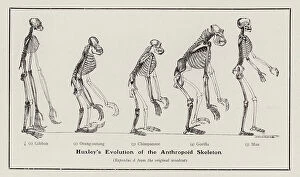
Background imageAnatomy Collection: Evolution of the human skeleton (litho)

Evolution of the human skeleton (litho)
8672117 Evolution of the human skeleton (litho) by English School, (19th century); Private Collection; (add.info.: Evolution of the human skeleton, as described by Thomas Henry Huxley)

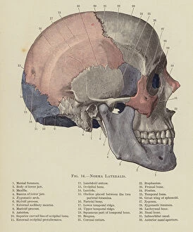
Background imageAnatomy Collection: Norma Lateralis (colour litho)

Norma Lateralis (colour litho)
8667626 Norma Lateralis (colour litho) by English School, (20th century); Private Collection; (add.info.: Norma Lateralis)

Background imageAnatomy Collection: Anterior View of Elbow Joint (colour litho)

Anterior View of Elbow Joint (colour litho)
8667622 Anterior View of Elbow Joint (colour litho) by English School, (20th century); Private Collection; (add.info.: Anterior View of Elbow Joint)

Background imageAnatomy Collection: Dissection of the Knee Joint from the Front, Patella thrown down (colour litho)

Dissection of the Knee Joint from the Front, Patella thrown down (colour litho)
8667620 Dissection of the Knee Joint from the Front, Patella thrown down (colour litho) by English School, (20th century); Private Collection; (add.info.: Dissection of the Knee Joint from the Front)

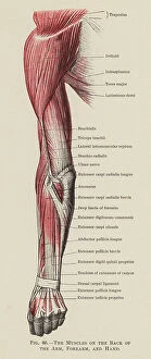
Background imageAnatomy Collection: The Muscles on the Back of the Arm, Forearm, and Hand (colour litho)

The Muscles on the Back of the Arm, Forearm, and Hand (colour litho)
8667619 The Muscles on the Back of the Arm, Forearm, and Hand (colour litho) by English School, (20th century); Private Collection; (add.info.: The Muscles on the Back of the Arm, Forearm, and Hand)

Background imageAnatomy Collection: Motor Points of the Upper Extremity, Anterior Aspect (litho)

Motor Points of the Upper Extremity, Anterior Aspect (litho)
8667614 Motor Points of the Upper Extremity, Anterior Aspect (litho) by English School, (20th century); Private Collection; (add.info.: Motor Points of the Upper Extremity, Anterior Aspect)

Background imageAnatomy Collection: Lynx lynx (Crane du lynx boreal ou lynx eurasian or wolf deer - Skull of an Eurasian lynx)

Lynx lynx (Crane du lynx boreal ou lynx eurasian or wolf deer - Skull of an Eurasian lynx)
CSO5006707 Lynx lynx (Crane du lynx boreal ou lynx eurasian or wolf deer - Skull of an Eurasian lynx), Museum d'histoire naturelle de Marseille; (add.info)

Background imageAnatomy Collection: engraving of the book of Ambroise Pare: Des Monsters et Prodiges (first edition 1573)

engraving of the book of Ambroise Pare: Des Monsters et Prodiges (first edition 1573)
PCT4260992 engraving of the book of Ambroise Pare: Des Monsters et Prodiges (first edition 1573, appendix: Monsters marine) - Triton et Sirene du Nile; (add.info)

Background imageAnatomy Collection: study of a body, hand, copperplate engraving, around 1700

study of a body, hand, copperplate engraving, around 1700
UTD4923684 study of a body, hand, copperplate engraving, around 1700; (add.info.: study of a body, hand, copperplate engraving)

Background imageAnatomy Collection: BARTHOLIN, Thomas (1616-1680). Danish physician, mathematician, and theologian

BARTHOLIN, Thomas (1616-1680). Danish physician, mathematician, and theologian
CUL4875538 BARTHOLIN, Thomas (1616-1680). Danish physician, mathematician, and theologian. He is best known for his work in the discovery of the lymphatic system in humans

Background imageAnatomy Collection: Human Anatomy book by MONTANA DE MONSERRATE, Bernardino, 1551 (engraving)

Human Anatomy book by MONTANA DE MONSERRATE, Bernardino, 1551 (engraving)
CUL4932051 Human Anatomy book by MONTANA DE MONSERRATE, Bernardino, 1551 (engraving) by Spanish School, (16th century); Biblioteca Nacional, Madrid, Spain; (add.info.: MONTANA DE MONSERRATE)

Background imageAnatomy Collection: Human Anatomy book by MONTANA DE MONSERRATE, Bernardino, 1551 (engraving)

Human Anatomy book by MONTANA DE MONSERRATE, Bernardino, 1551 (engraving)
CUL4932007 Human Anatomy book by MONTANA DE MONSERRATE, Bernardino, 1551 (engraving); Biblioteca Nacional, Madrid, Spain; (add.info.: MONTANA DE MONSERRATE, Bernardino (1480-1588). Spanish doctor

Background imageAnatomy Collection: Anatomical Plate - in ' De anatome corporis humani' by Guido Guidi (Vidus Vidius) (1509-1569)

Anatomical Plate - in " De anatome corporis humani" by Guido Guidi (Vidus Vidius) (1509-1569)
XEE5029671 Anatomical Plate - in " De anatome corporis humani" by Guido Guidi (Vidus Vidius) (1509-1569) Italian surgeon and anatomist

Background imageAnatomy Collection: Anatomical plate in ' De anatome corporis humani' by Guido Guidi (Vidus Vidius) (1509-1569)

Anatomical plate in " De anatome corporis humani" by Guido Guidi (Vidus Vidius) (1509-1569)
XEE5029670 Anatomical plate in " De anatome corporis humani" by Guido Guidi (Vidus Vidius) (1509-1569) Italian surgeon and anatomist

Background imageAnatomy Collection: Frontispice of ' De succo pancreatico', Leiden, 1671, by Regnier (Regnerus) de Graaf (1641 1673)

Frontispice of " De succo pancreatico", Leiden, 1671, by Regnier (Regnerus) de Graaf (1641 1673)
XEE5029587 Frontispice of " De succo pancreatico", Leiden, 1671, by Regnier (Regnerus) de Graaf (1641 1673) Dutch doctor - Engraving of 1672; (add.info)

Background imageAnatomy Collection: Monster creatures. Illustrated chart of a manuscript of Natural History by Ulisse Aldrovandi

Monster creatures. Illustrated chart of a manuscript of Natural History by Ulisse Aldrovandi (1522-1605)
VEN5029553 Monster creatures. Illustrated chart of a manuscript of Natural History by Ulisse Aldrovandi (1522-1605), Ulisse Aldrovandi fonds, University Library of Bologna, ed

Background imageAnatomy Collection: Monster creatures. Illustrated chart of a manuscript of Natural History by Ulisse Aldrovandi

Monster creatures. Illustrated chart of a manuscript of Natural History by Ulisse Aldrovandi (1522-1605)
VEN5029539 Monster creatures. Illustrated chart of a manuscript of Natural History by Ulisse Aldrovandi (1522-1605), Ulisse Aldrovandi fonds, University Library of Bologna, ed

Background imageAnatomy Collection: Monster creatures. Illustrated chart of a manuscript of Natural History by Ulisse Aldrovandi

Monster creatures. Illustrated chart of a manuscript of Natural History by Ulisse Aldrovandi (1522-1605)
VEN5029520 Monster creatures. Illustrated chart of a manuscript of Natural History by Ulisse Aldrovandi (1522-1605), Ulisse Aldrovandi fonds, University Library of Bologna, ed

Background imageAnatomy Collection: Cover page of ' Anatomia hepatis, cui praemittuntur quaedam ad rem anatomicam universe spectantia

Cover page of " Anatomia hepatis, cui praemittuntur quaedam ad rem anatomicam universe spectantia et ad calcem
XEE5029014 Cover page of " Anatomia hepatis, cui praemittuntur quaedam ad rem anatomicam universe spectantia et ad calcem operis subjiciuntur nonnulla de lymphae ductibus nuper repertis"

Background imageAnatomy Collection: Distribution of the hard portion of the auditory nerve - in ' Works Anatomiques'

Distribution of the hard portion of the auditory nerve - in " Works Anatomiques"
XEE5028966 Distribution of the hard portion of the auditory nerve - in " Works Anatomiques", Chez Charles-Antoine Jombert, 1761, Biblioteca nazionalez braidense

Background imageAnatomy Collection: Fossil skeleton of plesiosaurus by Lyme Regis. in ' Discours sur Les Revolutions de la Surface du

Fossil skeleton of plesiosaurus by Lyme Regis. in " Discours sur Les Revolutions de la Surface du Globe
XEE5028522 Fossil skeleton of plesiosaurus by Lyme Regis. in " Discours sur Les Revolutions de la Surface du Globe, et sur les changemens qu'elles ont produits dans Le Regne Anima" by

Background imageAnatomy Collection: Human skeleton found in Guadeloupe. in ' Discours sur Les Revolutions de La Surface du Globe

Human skeleton found in Guadeloupe. in " Discours sur Les Revolutions de La Surface du Globe
XEE5028515 Human skeleton found in Guadeloupe. in " Discours sur Les Revolutions de La Surface du Globe, et sur Les Changemens qu'elles produits dans Le Regne Animal " by Georges

Background imageAnatomy Collection: Anatomic plate in ' Anatomia Reformata' by Thomas Bartholin (1616-80) (Bartholinus)

Anatomic plate in " Anatomia Reformata" by Thomas Bartholin (1616-80) (Bartholinus) from 1651 - Venas Lacteas
XEE5027928 Anatomic plate in " Anatomia Reformata" by Thomas Bartholin (1616-80) (Bartholinus) from 1651 - Venas Lacteas; (add.info)


All Professionally Made to Order for Quick Shipping

"Unveiling the Intricacies of Anatomy: From Sensory Homunculus to Motor Homunculus" Step into the fascinating world of anatomy, where every detail tells a story. Explore the depths of human and animal structures as we delve into this captivating subject. Let's begin with Leonardo da Vinci's masterpiece, "Head of a Bear. " Created in 1480 but discovered in 1945, this artwork showcases da Vinci's unparalleled ability to capture anatomical accuracy. The intricate details reveal his keen observation skills and deep understanding of form. Moving on to our furry friends, the greyhound takes center stage. Discovering the anatomy of these majestic creatures unveils their remarkable speed and agility. From their sleek skeleton to their muscular build, it is evident why they are renowned for their racing abilities. But what about us humans? Enter the sensory homunculus – a visual representation that depicts how our brain perceives different body parts based on sensitivity levels. This map reveals intriguing insights into how our senses are distributed throughout our bodies. X-ray images provide another dimension to understanding anatomy. Take a glimpse at normal knees through an X-ray lens – marvel at the complexity hidden beneath our skin while appreciating its delicate balance between strength and flexibility. The backbone is often considered one of nature's most ingenious designs. Our human backbone includes ribs and pelvis, providing stability while allowing movement – truly an architectural marvel worth exploring further. Venturing deeper within ourselves, let us explore the intricacies of the human brain from an inferior view. Witness its complex network of connections responsible for controlling various bodily functions - truly awe-inspiring. As we journey back in time, we encounter hominid crania – remnants that shed light on our evolutionary history. These ancient skulls offer glimpses into early forms such as Australopithecus afarensis (AL 288-1), famously known as Lucy - bridging gaps between past and present.