Fraud Blocker Skip to main content

Anatomy Collection (page 9)

"Unveiling the Intricacies of Anatomy: From Sensory Homunculus to Motor Homunculus" Step into the fascinating world of anatomy, where every detail tells a story

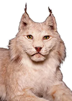
Background imageAnatomy Collection: Lynx sp. lynx

Lynx sp. lynx
Stuffed specimen from the collection of the Natural History Museum at Tring, part of the Natural History Museum, London

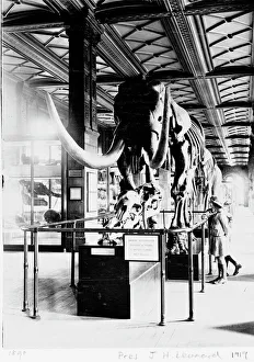
Background imageAnatomy Collection: Mastodon in Geological Gallery, December 1919

Mastodon in Geological Gallery, December 1919
The Guide Lecturer, John Henry Leonard, took this shot of two girls inspecting a primitive elephant or mastodon (Mammut americanus), in December 1919

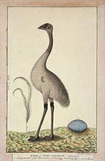
Background imageAnatomy Collection: Dromaius novaehollandiae, emu

Dromaius novaehollandiae, emu
Drawing No. 67, watercolour by George Raper (1792) from The Raper Collection

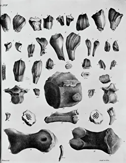
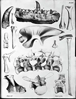
Background imageAnatomy Collection: Plate 17. Mantells Geology of Sussex

Plate 17. Mantells Geology of Sussex
Plate 17 from Illustrations of the Geology of Sussex, 1827 by G. A. Mantell

Background imageAnatomy Collection: Plate 19. Mantells Geology of Sussex

Plate 19. Mantells Geology of Sussex
Plate 19 from Illustrations of the Geology of Sussex, 1827 by G. A. Mantell

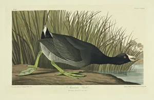
Background imageAnatomy Collection: Fulica americana, American coot

Fulica americana, American coot
Plate 239 from John James Audubons Birds of America, original double elephant folio (1834-35), hand-coloured aquatint. Engraved, printed and coloured by R. Havell (& Son), London

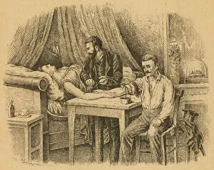
Background imageAnatomy Collection: Transfusing Blood, 1880

Transfusing Blood, 1880
TRANSFUSION - a doctor takes blood from the husband to give to the wife

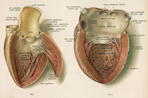
Background imageAnatomy Collection: Inside the Heart

Inside the Heart
No man knoweth the secrets of the heart - but these interior views give you some idea... Date: late 19th century

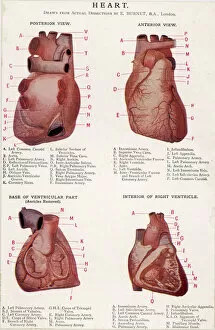
Background imageAnatomy Collection: 4 Views of Heart

4 Views of Heart
Four views of the heart, posterior and anterior, and the base and interior of the ventricle

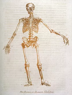
Background imageAnatomy Collection: Anatomy: human Skeleton

Anatomy: human Skeleton
The human skeleton

Background imageAnatomy Collection: Saffron Crocus - source of saffron. (Stigmas used) Greece

Saffron Crocus - source of saffron. (Stigmas used) Greece
ROG-10908 Saffron Crocus - source of saffron. (Stigmas used) Greece Crocus sativus Bob Gibbons Please note that prints are for personal display purposes only and may not be reproduced in anyway

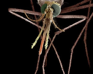
Background imageAnatomy Collection: Scanning Electron Micrograph (SEM): Mosquito, Female; Magnification x 55 (A4 size: 29. 7 cm width)

Scanning Electron Micrograph (SEM): Mosquito, Female; Magnification x 55 (A4 size: 29. 7 cm width)
LRDS-83 Mosquito, Female Scanning Electron Micrograph (SEM) Anopheles sp. Magnification x 55 (A4 size: 29.7 cm width) Coloured by hand to enhance natural features

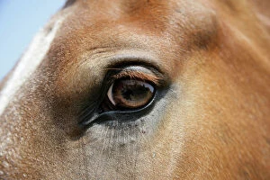
Background imageAnatomy Collection: Horse. Eye

Horse. Eye
JD-19379 Horse. Eye John Daniels Please note that prints are for personal display purposes only and may not be reproduced in any way. contact details: prints@ardea.com tel: + 44 (0) 20 8318 1401

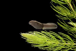
Background imageAnatomy Collection: Common toad – tadpole 5 weeks – x0. 6 – black background Bedfordshire UK 004542

Common toad – tadpole 5 weeks – x0. 6 – black background Bedfordshire UK 004542
BB-1244 Common toad - tadpole 5 weeks, x0.6 Bedfordshire, UK Bufo bufo Brian Bevan Please note that prints are for personal display purposes only and may not be reproduced in any way

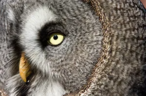
Background imageAnatomy Collection: Great grey owl - Close-up of face. Adult

Great grey owl - Close-up of face. Adult
VG-22983 Great grey owl - Close-up of face. Adult Strix nebulosa Distribution: Northern Europe, northern Asia and northern parts of United States and Canada

Background imageAnatomy Collection: Hybrid Orchid - detailed study of blossom, arboreal type. Lower Saxony, Germany

Hybrid Orchid - detailed study of blossom, arboreal type. Lower Saxony, Germany
USH-2586 Hybrid Orchid - detailed study of blossom, arboreal type Lower Saxony, Germany Phalaenopsis Mystik isles Duncan Usher Please note that prints are for personal display purposes only

Background imageAnatomy Collection: Swallowtail Butterfly - on blackthorn

Swallowtail Butterfly - on blackthorn
VG-227 Swallowtail Butterfly - on blackthorn Papillio machaon Paul Van Gaalen Please note that prints are for personal display purposes only and may not be reproduced in any way

Background imageAnatomy Collection: Dog - Airdale terrier

Dog - Airdale terrier
LA-1514 Dog - Airedale TERRIER - close-up of face Jean Michel Labat Please note that prints are for personal display purposes only and may not be reproduced in any way

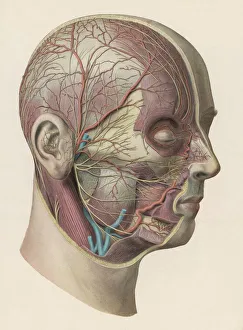
Background imageAnatomy Collection: Diagram - Inside of Head

Diagram - Inside of Head
Detailed diagram showing muscles and veins inside of the head

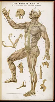
Background imageAnatomy Collection: Muscles of Human Body

Muscles of Human Body
Physiological diagram of the muscles, joints and animal mechanics of the human body

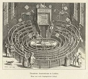
Background imageAnatomy Collection: Anatomy Theatre, Leiden

Anatomy Theatre, Leiden
THE ANATOMY THEATRE at LEIDEN, Netherlands

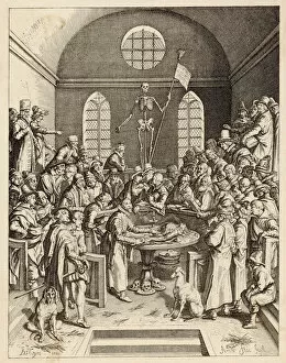
Background imageAnatomy Collection: Medical / Anatomy C16

Medical / Anatomy C16
Anatomy demonstration. Date: 16th Century

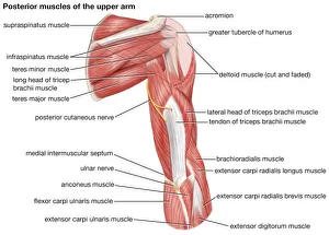
Background imageAnatomy Collection: A posterior view of the muscles of the human upper arm

A posterior view of the muscles of the human upper arm

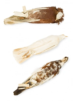
Background imageAnatomy Collection: Darwins Pigeons

Darwins Pigeons
Charles Darwin gave his personal collection on pigeons to the Museum in 1867 and 1868, as part of a bigger collection of domestic birds including ducks, chickens and even cararies

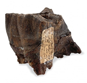
Background imageAnatomy Collection: Tooth from Coelodonta antiquitatis, woolly rhinoceros

Tooth from Coelodonta antiquitatis, woolly rhinoceros
This woolly rhino tooth, together with two others were found in 1668 at Chartham, near Canterbury in Kent by natural historian John Somner

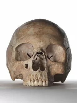
Background imageAnatomy Collection: Modern human skull

Modern human skull
Skull of an adult, female modern human

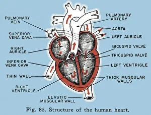
Background imageAnatomy Collection: Labelled Structure of the Human Heart Diagram

Labelled Structure of the Human Heart Diagram
http://csaimages.com/images/istockprofile/csa_vector_dsp.jpg

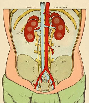
Background imageAnatomy Collection: Anatomy of Body Featuring Kidneys

Anatomy of Body Featuring Kidneys
http://csaimages.com/images/istockprofile/csa_vector_dsp.jpg

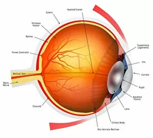
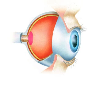
Background imageAnatomy Collection: Human eye, illustration

Human eye, illustration
Eye anatomy. Cutaway illustration passing through a human eye, showing its internal anatomy and structure. The front of the eye is at right, and the structures here include the cornea

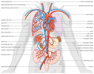
Background imageAnatomy Collection: Human arterial supply and venous drainage of the organs

Human arterial supply and venous drainage of the organs

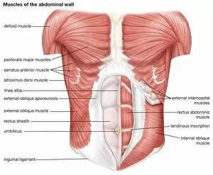
Background imageAnatomy Collection: The muscles of the human abdominal wall

The muscles of the human abdominal wall

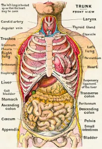
Background imageAnatomy Collection: Anatomical View of the Human Torso, 1914 (engraving)

Anatomical View of the Human Torso, 1914 (engraving)
2622096 Anatomical View of the Human Torso, 1914 (engraving) by German School, (20th century); Private Collection; (add.info.: Vintage anatomical study of the human torso, frontal view)

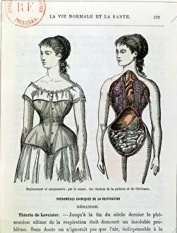
Background imageAnatomy Collection: The Harmful Effects of the Corset, illustration from La Vie Normale et la Sante

The Harmful Effects of the Corset, illustration from La Vie Normale et la Sante
CHT171318 The Harmful Effects of the Corset, illustration from La Vie Normale et la Sante by Dr Jules Rengade (b.1841) c.1880 (colour litho) by French School, (19th century); Bibliotheque Nationale

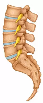
Background imageAnatomy Collection: Normal lateral view of the lumbar vertebrae showing spinal nerve roots

Normal lateral view of the lumbar vertebrae showing spinal nerve roots
Detail, Anatomy, Diagram, Illustration, Spine, Bone, Medical, Biology, Disk, Normal, Cut Out, White Background, Vertical, Artwork, Color Image, Close Up, Healthcare And Medicine, The Human Body

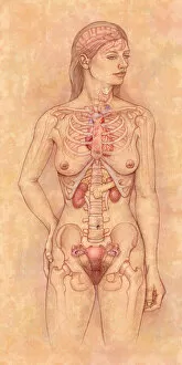
Background imageAnatomy Collection: Front view of the female anatomy hilighting the endocrine system

Front view of the female anatomy hilighting the endocrine system
Brain, Anatomy, Diagram, Heart, Illustration, Bone, Shoulder, Medical, Uterus, Biology, Pelvis, Femur, See Through, Vertical, Artwork, Female Likeness, Color Image, Front View

Background imageAnatomy Collection: Human brain, illustration

Human brain, illustration
Illustration of a cross-section of the brain showing the various lobes. The lobes are shown in different colours - red (frontal), green (parietal), yellow (occipital), orange (temporal)

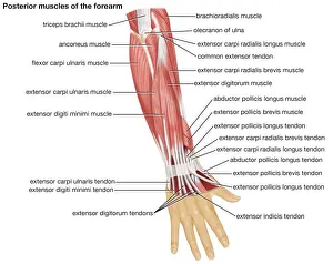
Background imageAnatomy Collection: The posterior view of the muscles of the human forearm

The posterior view of the muscles of the human forearm

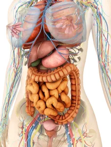
Background imageAnatomy Collection: Female body showing digestive and circulatory system

Female body showing digestive and circulatory system
Medical illustration of female body showing digestive and circulatory system, perspective view

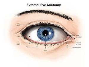
Background imageAnatomy Collection: External anatomy of the human eye (with labels)

External anatomy of the human eye (with labels)

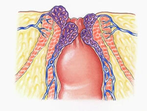
Background imageAnatomy Collection: Cross section illustration of human anal column showing external and internal sphincters

Cross section illustration of human anal column showing external and internal sphincters, healthy veins
Anal, Anatomy, Anus, Biomedical Illustration, Blood Flow, Close-Up, Column, Cross Section, External, Haemorrhoids, Healthcare and Medicine, Healthy, Human Vein, Inflammation, Internal, No People

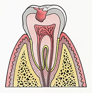
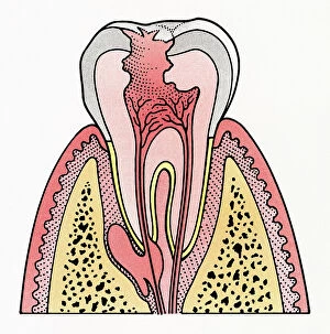
Background imageAnatomy Collection: Cross section illustration showing tooth decay

Cross section illustration showing tooth decay
Anatomy, Bacterium, Biomedical Illustration, Blood Supply, Close-Up, Cross Section, Decay, Dental Hygiene, Enamel, Full Length, Healthcare and Medicine, Human Bone, Human Jaw Bone, Human Teeth

Background imageAnatomy Collection: Cross section illustration of anatomy of human eye showing use of optic nerve

Cross section illustration of anatomy of human eye showing use of optic nerve
Anatomy, Biomedical Illustration, Close-Up, Cross Section, Eyelash, Fovea, Human Eye, Iris, No People, Optic Nerve, Part Of, Physiology, Pupil, Sensory Perception, Side View, Single Object

Background imageAnatomy Collection: Cross section illustration showing tooth decay

Cross section illustration showing tooth decay
Anatomy, Bacterium, Biomedical Illustration, Blood Supply, Close-Up, Cross Section, Decay, Dental Hygiene, Enamel, Full Length, Healthcare and Medicine, Human Bone, Human Jaw Bone, Human Teeth

Background imageAnatomy Collection: A damaged bronchus

A damaged bronchus
Anatomy, Biomedical Illustration, Bronchitis, Bronchus, Chronic, Close-Up, Damaged, Healthcare and Medicine, Illness, No People, Pen and Ink, Problems, Watercolour Painting, White Background

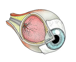
Background imageAnatomy Collection: Cross-section of human eye

Cross-section of human eye
Anatomy, Cross-Section, Eyeball, Eyesight, Human Eye, Nerve, No People, Pen and Ink, Physiology, Single Object, Watercolour Painting, White Background, Horizontal, Dorling Kindersley Prints, 88008085

Background imageAnatomy Collection: Muscles in human hand and arm

Muscles in human hand and arm
Anatomy, Biomedical Illustration, Close-Up, Healthcare and Medicine, Human Arm, Human Hand, Human Muscle, Human Representation, No People, White Background, Tendons, Watercolour Painting

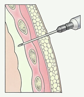
Background imageAnatomy Collection: Syringe entering pleural membranes around human lung to extract sample

Syringe entering pleural membranes around human lung to extract sample
Anatomy, Biomedical Illustration, Close-Up, Cross-Section, Extracting, Healthcare and Medicine, Human Lung, Medical Instrument, Medical Procedure, Medical Sample, Operation, Pen and Ink


All Professionally Made to Order for Quick Shipping

"Unveiling the Intricacies of Anatomy: From Sensory Homunculus to Motor Homunculus" Step into the fascinating world of anatomy, where every detail tells a story. Explore the depths of human and animal structures as we delve into this captivating subject. Let's begin with Leonardo da Vinci's masterpiece, "Head of a Bear. " Created in 1480 but discovered in 1945, this artwork showcases da Vinci's unparalleled ability to capture anatomical accuracy. The intricate details reveal his keen observation skills and deep understanding of form. Moving on to our furry friends, the greyhound takes center stage. Discovering the anatomy of these majestic creatures unveils their remarkable speed and agility. From their sleek skeleton to their muscular build, it is evident why they are renowned for their racing abilities. But what about us humans? Enter the sensory homunculus – a visual representation that depicts how our brain perceives different body parts based on sensitivity levels. This map reveals intriguing insights into how our senses are distributed throughout our bodies. X-ray images provide another dimension to understanding anatomy. Take a glimpse at normal knees through an X-ray lens – marvel at the complexity hidden beneath our skin while appreciating its delicate balance between strength and flexibility. The backbone is often considered one of nature's most ingenious designs. Our human backbone includes ribs and pelvis, providing stability while allowing movement – truly an architectural marvel worth exploring further. Venturing deeper within ourselves, let us explore the intricacies of the human brain from an inferior view. Witness its complex network of connections responsible for controlling various bodily functions - truly awe-inspiring. As we journey back in time, we encounter hominid crania – remnants that shed light on our evolutionary history. These ancient skulls offer glimpses into early forms such as Australopithecus afarensis (AL 288-1), famously known as Lucy - bridging gaps between past and present.