Framed Print : Light micrograph of normal heart (cardiac) muscle
 muscle")
 muscle")

![]()
Framed Photos from Science Photo Library
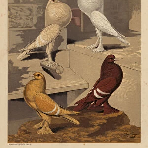
Light micrograph of normal heart (cardiac) muscle
Heart muscle. Light micrograph of healthy heart (cardiac) muscle, seen in longitudinal section. Bundles of muscle fibres are stained red, running diagonally from upper right to lower left. Fibres are joined end to end by intercalated discs (not visible), forming a continuous network which allows for efficient tranmission and regulation of muscle contraction. Fibre bundles are joined across spaces (white) by connective tissue. This connective tissue also occurs between muscle cells and is extremely rich in blood capillaries. The high concentration of blood vessels in heart muscle reflects its demand for oxygen and high performance. Magnification: x125 at 6x7cm size
Science Photo Library features Science and Medical images including photos and illustrations
Media ID 6420298
© PASIEKA/SCIENCE PHOTO LIBRARY
Cardiac Cardiac Muscle Heart Muscle Muscle Fibre Muscles
12"x10" Modern Frame
Discover the intricacy of life with our Media Storehouse Framed Prints featuring this stunning light micrograph of a healthy heart (cardiac) muscle. Captured by Science Photo Library, this image showcases the beauty of bundles of muscle fibers, stained red, running diagonally from upper right to lower left. A perfect addition to any home or office space, this framed print brings the wonders of science into your daily life. Explore our range of scientific and artistic prints, and let the beauty of knowledge inspire you.
10x8 Print in an MDF Wooden Frame with 180 gsm Satin Finish Paper. Glazed using shatter proof thin plexi glass. Frame thickness is 1 inch and depth 0.75 inch. Fluted cardboard backing held with clips. Supplied ready to hang with sawtooth hanger and rubber bumpers. Spot clean with a damp cloth. Packaged foam wrapped in a card.
Contemporary Framed and Mounted Prints - Professionally Made and Ready to Hang
Estimated Image Size (if not cropped) is 25.4cm x 25.4cm (10" x 10")
Estimated Product Size is 25.4cm x 30.5cm (10" x 12")
These are individually made so all sizes are approximate
Artwork printed orientated as per the preview above, with landscape (horizontal) or portrait (vertical) orientation to match the source image.
EDITORS COMMENTS
This print showcases a light micrograph of normal heart (cardiac) muscle, providing an intricate glimpse into the inner workings of our vital organ. The image captures a longitudinal section of healthy heart muscle, revealing bundles of vibrant red-stained muscle fibers that run diagonally from the upper right to the lower left. What makes this microscopic view even more fascinating is the presence of intercalated discs, although not visible in this particular image. These discs serve as connectors between individual muscle fibers, forming a continuous network crucial for efficient transmission and regulation of muscle contraction. The connective tissue depicted here acts as both a bridge across spaces and a rich source of blood capillaries. This abundance of blood vessels within the heart muscle signifies its constant need for oxygen and exceptional performance capabilities. With a magnification level set at x125 on a 6x7cm size scale, this remarkable photograph offers us an extraordinary insight into the complex anatomy and functionality behind cardiac muscles. It serves as a testament to Science Photo Library's commitment to capturing awe-inspiring images that deepen our understanding of the human body without any commercial intentions involved.
MADE IN THE USA
Safe Shipping with 30 Day Money Back Guarantee
FREE PERSONALISATION*
We are proud to offer a range of customisation features including Personalised Captions, Color Filters and Picture Zoom Tools
SECURE PAYMENTS
We happily accept a wide range of payment options so you can pay for the things you need in the way that is most convenient for you
* Options may vary by product and licensing agreement. Zoomed Pictures can be adjusted in the Cart.