

Framed Prints From Alinari
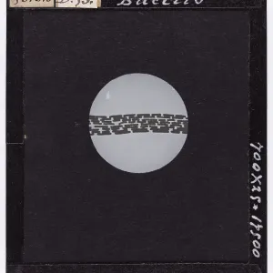
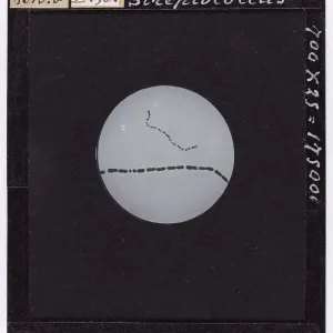
Framed Print of Negri bodies present in the cell of the of the Horn of Ammon of a rabid cow, enlarged under a microscope
Negri bodies present in the cell of the of the Horn of Ammon of a rabid cow, enlarged under a microscope. Date of Photograph:1900-1915 ca. © Alinari Archives, Florence
Artwork Licensed From Alinari
Your chosen combination is quickly and professionally made to order, our shipping page has more details
If you are not entirely happy with your items, simply return them to us within 30 days for a replacement or full refund. Please read our full returns policy.
Media ID 33224175
Editors Comments
Discover the fascinating world of microbiology with Media Storehouse's Framed Prints featuring the iconic image "Negri Bodies in the Cell of the Horn of Ammon of a Rabid Cow, Enlarged under a Microscope" by Giorgio Roster. This photograph, taken between 1900-1915, offers a glimpse into the intricacies of medical history. The print shows the distinct Negri bodies, a characteristic feature of rabies infection, magnified under the microscope. Add this educational piece to your home or office decor, and bring a touch of scientific discovery to your space.
This artwork is also available as...
Photo Prints - Discover the fascinating world of microbiology with Media Storehouse's photographic print by renowned Italian photographer Giorgio Roster. This image shows Negri bodies, named after Camillo Negri who first discovered them, in the cell of a rabid cow's Horn of Ammon. These distinctive structures are hallmarks of rabies infection and were captured under a microscope during this time period around 1900-1915. Bring the intrigue of scientific discovery into your home or office with this beautifully preserved, museum-quality print.
Poster Prints - Discover the intriguing world of microbiology with Media Storehouse's poster prints. This image, "Negri Bodies in the Cell of the Horn of Ammon of a Rabid Cow, Enlarged under a Microscope," is a rare find from the archive of renowned Italian photographer Giorgio Roster. Dating back to the early 1900s, this photograph shows the distinctive Negri bodies, which were among the first morphological markers identified for rabies virus infection. Bring this fascinating piece of scientific history into your home or office, and elevate your space with Media Storehouse's high-quality, poster prints.
Metal Prints - Discover the intriguing world of microbiology with Media Storehouse's Metal Prints featuring the iconic image "Negri Bodies in the Cell of the Horn of Ammon of a Rabid Cow," captured by renowned photographer Giorgio Roster in the early 1900s. This print shows the enlarged image of Negri bodies, a key discovery in the study of rabies, as they appear under a microscope. Add this educational piece to your decor, and bring the wonders of science into your home or office.
Canvas Prints - Discover the intriguing world of microbiology with Media Storehouse's Canvas Prints featuring the iconic image "Negri Bodies in the Cell of the Horn of Ammon of a Rabid Cow," enlarged under a microscope, by renowned photographer Giorgio Roster. This image, taken between 1900-1915, shows the distinctive Negri bodies, key indicators of rabies infection, found within an Ammon's horn cell. Bring this fascinating piece of scientific history into your home or office and ignite conversations with its stunning, canvas print. Experience the beauty of science like never before with Media Storehouse.
Modern Frame 12"x10"
Ready to Hang Contemporary Framed Prints adds style, protection, and a polished touch to your space - effortlessly.
Your artwork will be printed as shown in the preview. To adjust please select Edit Printed Image under the Buy button
Made and Shipped from within the USA or Canada as appropriate. Printed on 10"x8" Premium Matte Paper offering bright, white paper with a matte finish with a digitally rendered mat surround offering the timeless look of professionally mounted artwork. Our 1″ Frames are available in a range of colours with clear acrylic shatterproof glazing and Kraft pad backer. Matte Paper offer a lighter-weight alternative to fine art paper which is optimized for artwork and can be suitable for photography in scenarios where a less glossy appearance is preferred. This sustainable paper printing is an alternative to our premium fine art paper. Each frame is 1″ wide by 3/4″ deep and glazed using standard 1/16” clear PETG to protect and enhance the visibility of each print. Ready to hang with a sawtooth metal hanger and sturdy backer.
These are individually made so all sizes are approximate
Estimated Image Size (if not cropped) is 15.3cm x 16.9cm (6" x 6.7")
Estimated Product Size is 25.4cm x 30.5cm (10" x 12")