Canvas Print : Human foot musculature, artwork F007 / 3242


![]()
Canvas Prints From Science Photo Library
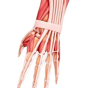
Human foot musculature, artwork F007 / 3242
Human foot musculature, computer artwork
Science Photo Library features Science and Medical images including photos and illustrations
Media ID 9272225
© PIXOLOGICSTUDIO/SCIENCE PHOTO LIBRARY
Extensor Digitorum Brevis Extensor Digitorum Longus Extensor Hallucis Brevis Extensor Hallucis Longus Foot High Angle View Human Body Part Human Foot Human Leg Joint Limb Metatarsal Peroneus Brevis Phalanx Tarsal Tendon Tibia Tibialis Anterior Human Skeleton Musculature
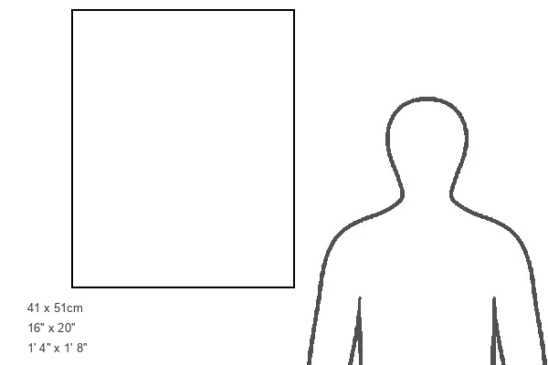
20"x16" (51x41cm) Canvas Print
Bring the intricacies of the human body into your home or office with our stunning Media Storehouse Canvas Prints. This particular piece, titled "Human foot musculature, artwork F007 / 3242" by PIXOLOGICSTUDIO/SCIENCE PHOTO LIBRARY, showcases a captivating computer-generated representation of the complex foot musculature. The vibrant colors and crisp details of this artwork are sure to impress, making it a perfect conversation starter and a unique addition to any space. Order your Media Storehouse Canvas Print today and bring the wonders of science into your home or workplace.
Delivered stretched and ready to hang our premium quality canvas prints are made from a polyester/cotton blend canvas and stretched over a 1.25" (32mm) kiln dried knot free wood stretcher bar. Packaged in a plastic bag and secured to a cardboard insert for safe transit.
Canvas Prints add colour, depth and texture to any space. Professionally Stretched Canvas over a hidden Wooden Box Frame and Ready to Hang
Estimated Product Size is 40.6cm x 50.8cm (16" x 20")
These are individually made so all sizes are approximate
Artwork printed orientated as per the preview above, with portrait (vertical) orientation to match the source image.
EDITORS COMMENTS
This print showcases the intricate musculature of a human foot, beautifully depicted through computer artwork. Against a striking black background, this illustration provides a detailed front view of an adult foot, offering insights into its biology and anatomy. The image highlights the various muscles, joints, and bones that make up this essential part of our body. From the tibia to the tarsal and metatarsal bones, every detail is meticulously portrayed. The phalanx bones are also visible, along with tendons that play a crucial role in movement. With its emphasis on healthy anatomy and normal structure, this artwork serves as an invaluable resource for studying human foot musculature. Its high angle view allows us to appreciate the complexity of these structures from a unique perspective. Notably featured are key muscles such as the tibialis anterior and peroneus brevis alongside extensor digitorum brevis and extensor hallucis brevis. Additionally, prominent longus muscles like extensor digitorum longus and extensor hallucis longus are showcased in remarkable detail. This visually stunning representation captures both scientific accuracy and artistic finesse. It offers viewers an opportunity to delve into the intricacies of our own bodies while appreciating the beauty found within anatomical structures.
MADE IN THE USA
Safe Shipping with 30 Day Money Back Guarantee
FREE PERSONALISATION*
We are proud to offer a range of customisation features including Personalised Captions, Color Filters and Picture Zoom Tools
SECURE PAYMENTS
We happily accept a wide range of payment options so you can pay for the things you need in the way that is most convenient for you
* Options may vary by product and licensing agreement. Zoomed Pictures can be adjusted in the Basket.