Home > Popular Themes > Red Arrows
Inversion of the foot, artwork C016 / 6802

![]()
Wall Art and Photo Gifts from Science Photo Library
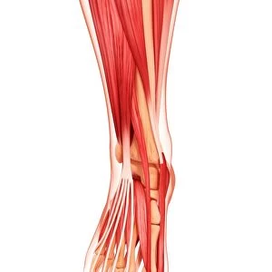
Inversion of the foot, artwork C016 / 6802
Inversion of the foot. Artwork of the muscles of the foot from above, with red arrows showing the direction of movement when inverting the foot. This is the motion that tilts the foot sideways so that the sole moves towards the midplane of the body. The opposite movement is eversion. Inversion is most commonly encountered when twisting the ankle. The muscles involved are the tibialis anterior and tibialis posterior. The nerve used is the tibial nerve. This is the left foot. For the right foot, see C016/6801
Science Photo Library features Science and Medical images including photos and illustrations
Media ID 9246157
© D & L GRAPHICS / SCIENCE PHOTO LIBRARY
Ankle Arthrology Biomechanics Diagram Foot From Above Joint Ligament Ligaments Limb Movement Moving Muscles Muscular Physiological Physiology Range Of Movements Superior Tendon Tendons Tibial Nerve Tibialis Anterior Tibialis Posterior Twist Twisting Cutouts Inversion Invert Left Foot Musculature
FEATURES IN THESE COLLECTIONS
EDITORS COMMENTS
This photo print, titled "Inversion of the foot, artwork C016 / 6802" offers a detailed illustration of the muscles and movements involved in tilting the foot sideways towards the midplane of the body. Against a pristine white background, this anatomical masterpiece showcases the intricate biology and physiology behind this motion. Highlighted by vibrant red arrows, which indicate the direction of movement during inversion, this artwork focuses on one specific aspect: twisting the ankle. Inversion is commonly encountered when subjecting our ankles to such twists. The key players in this action are two vital muscles - tibialis anterior and tibialis posterior - both controlled by the tibial nerve. With its cut-out presentation from above, this image provides an exceptional view into not only these muscles but also various other components that contribute to foot movement. Ligaments, tendons, and joints all play their part in ensuring a healthy range of movements for our feet. Created by D & L GRAPHICS for Science Photo Library's collection, this print serves as an invaluable resource for those interested in studying biomechanics or understanding human anatomy at a deeper level. Its scientific precision combined with artistic flair makes it an ideal addition to any educational setting or medical facility seeking to explore musculature and joint mechanics within our remarkable bodies.
MADE IN THE USA
Safe Shipping with 30 Day Money Back Guarantee
FREE PERSONALISATION*
We are proud to offer a range of customisation features including Personalised Captions, Color Filters and Picture Zoom Tools
SECURE PAYMENTS
We happily accept a wide range of payment options so you can pay for the things you need in the way that is most convenient for you
* Options may vary by product and licensing agreement. Zoomed Pictures can be adjusted in the Cart.