Fraud Blocker Skip to main content

Ligaments Collection

"Ligaments: The Unsung Heroes of Our Body's Stability and Mobility" Damaged knee ligament, artwork: A visual representation of the intricate network in our knees

Background imageLigaments Collection: Damaged knee ligament, artwork

Damaged knee ligament, artwork
Damaged knee ligament. Computer artwork of the muscle structure of the leg and anatomy of the human knee joint, showing a tear (dark line, centre right) in one of the cruciate ligaments (beige)

Background imageLigaments Collection: Skeleton & Ligaments

Skeleton & Ligaments
Physiological diagram of the skeleton and ligaments

Background imageLigaments Collection: Arm circulation, anatomical artwork C013 / 7419

Arm circulation, anatomical artwork C013 / 7419
Arm anatomy. Computer artwork showing the muscles (brown), ligaments (white), arteries (red), veins (blue), lymph vessels (yellow), and lymph nodes (round, yellow) of a human arm

Background imageLigaments Collection: Muscles of the neck

Muscles of the neck, historical artwork. The upper figure shows the front of the neck with the skin and fascia (connective tissue) removed to expose the musculature (red) and tendons

Background imageLigaments Collection: Outer ankle ligaments, artwork C013 / 4452

Outer ankle ligaments, artwork C013 / 4452
Outer ankle ligaments. Computer artwork of the bones and ligaments (white) of the right foot and ankle seen from the side

Background imageLigaments Collection: Eye muscle, TEM C014 / 1468

Eye muscle, TEM C014 / 1468
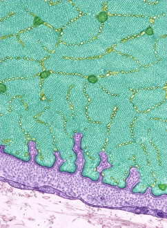
Eye muscle. Transmission electron micrograph (TEM) of a section through a striated muscle cell from the ciliary muscle of a human eye

Background imageLigaments Collection: Inner ankle ligaments, artwork C013 / 4451

Inner ankle ligaments, artwork C013 / 4451
Inner ankle ligaments. Computer artwork of the bones and ligaments (white) of the right foot and ankle seen from the side

Background imageLigaments Collection: Muscles (engraving)

Muscles (engraving)
471461 Muscles (engraving) by Rubens, Peter Paul (1577-1640); 34.4x24 cm; Koninklijk Museum voor Schone Kunsten, Antwerp, Belgium; © Art in Flanders

Background imageLigaments Collection: Ligaments of the human body (engraving)

Ligaments of the human body (engraving)
5216894 Ligaments of the human body (engraving) by German School, (19th century); Private Collection; (add.info.: Ligaments of the human body)

Background imageLigaments Collection: Study of the Muscles of the Human Torso and Neck, 1906 (engraving)

Study of the Muscles of the Human Torso and Neck, 1906 (engraving)
2622227 Study of the Muscles of the Human Torso and Neck, 1906 (engraving) by German School, (20th century); Private Collection; (add.info)

Background imageLigaments Collection: Hand muscle anatomy, 1831 artwork

Hand muscle anatomy, 1831 artwork
Hand muscle anatomy. Radial view of the ligaments and muscles of the hand (main artwork, centre). The insets show various aspects of the ligaments, muscles, and bones of the fingers

Background imageLigaments Collection: Muscles of the forearm, 1831 artwork

Muscles of the forearm, 1831 artwork
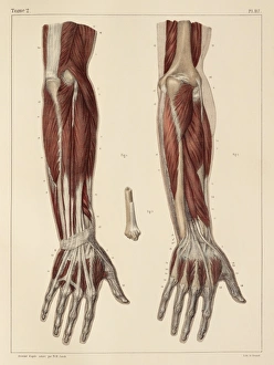
Muscles of the forearm, posterior view. The inset shows the inferior attachment of the long supinator muscle. This anatomical artwork is plate 117 from volume 2 (1831)

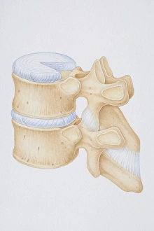
Background imageLigaments Collection: Diagram of a human thoracic vertebrae including the intervertebral disk

Diagram of a human thoracic vertebrae including the intervertebral disk, vertebral process and ligaments

Background imageLigaments Collection: Diagram illustrating the human musculo-skeletal system

Diagram illustrating the human musculo-skeletal system

Background imageLigaments Collection: Cross-section diagram of human foot, side view

Cross-section diagram of human foot, side view

Background imageLigaments Collection: Human thorax, shoulder girdle, and pelvis posterior view

Human thorax, shoulder girdle, and pelvis posterior view

Background imageLigaments Collection: Fist clenching movement, artwork C016 / 6795

Fist clenching movement, artwork C016 / 6795
Fist clenching movement. Artwork of the muscles of the forearm and hand from the front, with red arrows showing the direction of movement of the fingers during flexion to form a fist

Background imageLigaments Collection: Little and ring finger flexion, artwork C016 / 6793

Little and ring finger flexion, artwork C016 / 6793
Little and ring finger flexion. Artwork of the muscles of the forearm and hand from the front, with red arrows showing the direction of movement of the little and ring (fourth and third)

Background imageLigaments Collection: Arm anatomy, artwork C013 / 4583

Arm anatomy, artwork C013 / 4583
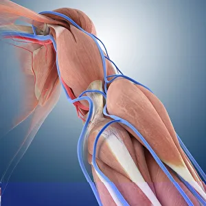
Arm anatomy. Computer artwork of an oblique anterior view looking up a human arm, showing muscles and tendons (brown and white), bones and ligaments (white), arteries and veins (red and blue)

Background imageLigaments Collection: Buttock muscles, artwork C013 / 4414

Buttock muscles, artwork C013 / 4414
Buttock muscles. Computer artwork of a posterior view of the hips showing some of the muscles (red) of the buttocks. These are shown attached to the femurs (thigh bones) and the pelvis bones

Background imageLigaments Collection: Foot anatomy

Foot anatomy, computer artwork. Muscles are pink, tendons and ligaments are white and bones are cream. Tendons and ligaments are bands of inelastic fibrous tissue

Background imageLigaments Collection: Foot bones and ligaments

Foot bones and ligaments
Antique illustration of a foot bones and ligaments

Background imageLigaments Collection: Human foot, internal anatomy

Human foot, internal anatomy

Background imageLigaments Collection: Knee anatomy, artwork F008 / 3210

Knee anatomy, artwork F008 / 3210
Knee anatomy, artwork

Background imageLigaments Collection: Human foot anatomy, artwork

Human foot anatomy, artwork
Human foot anatomy, computer artwork

Background imageLigaments Collection: Lower leg muscles, 1831 artwork

Lower leg muscles, 1831 artwork
Lower leg muscles. This anatomical artwork is plate 141 from volume 2 (1831) of Traite complet de l anatomie de l homme (1831-1854)

Background imageLigaments Collection: Plantar foot muscles, 1831 artwork

Plantar foot muscles, 1831 artwork
Plantar foot muscles. This anatomical artwork is plate 144 from volume 2 (1831) of Traite complet de l anatomie de l homme (1831-1854)

Background imageLigaments Collection: Inversion of the foot, artwork C016 / 6802

Inversion of the foot, artwork C016 / 6802
Inversion of the foot. Artwork of the muscles of the foot from above, with red arrows showing the direction of movement when inverting the foot

Background imageLigaments Collection: Foot ligaments and tendons, artwork C016 / 7010

Foot ligaments and tendons, artwork C016 / 7010
Foot ligaments and tendons. Artwork of the bones, ligaments (white, between bones), and tendons (white, muscle-bone) of the foot and the ankle joint from the side

Background imageLigaments Collection: Elbow flexion, artwork C016 / 6790

Elbow flexion, artwork C016 / 6790
Elbow flexion. Artwork of the muscles of the arm from the front, with red arrows showing the direction of movement when flexing (bending) the elbow

Background imageLigaments Collection: Knee muscles and bones, artwork C016 / 7013

Knee muscles and bones, artwork C016 / 7013
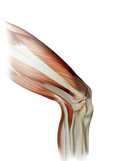
Knee muscles and bones. Artwork of an oblique lateral frontal view of the muscles and bones of a human knee. This view is of the outside of the left leg

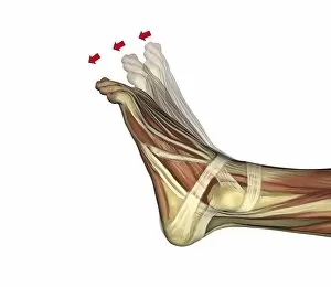
Background imageLigaments Collection: Plantarflexion of the foot, artwork C016 / 6800

Plantarflexion of the foot, artwork C016 / 6800
Plantarflexion of the foot. Artwork of the muscles of the foot from the side, with red arrows showing the direction of movement when flexing the foot in the direction of its lower (plantar)

Background imageLigaments Collection: Atlanto-occipital joint, artwork C016 / 6554

Atlanto-occipital joint, artwork C016 / 6554
Atlanto-occipital joint. Artwork of the bones (yellow) and ligaments (blue) of the atlanto-occipital joint, as seen from behind (posterior view)

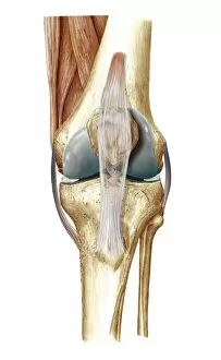
Background imageLigaments Collection: Knee bones and ligaments, artwork C016 / 7012

Knee bones and ligaments, artwork C016 / 7012
Knee bones and ligaments. Artwork of a frontal view of the knee joint, showing bones, ligaments, tendons, and cartilage. The bones are the femur (thigh bone, top), patella (knee-cap, upper centre)

Background imageLigaments Collection: Elbow flexion, artwork C016 / 6789

Elbow flexion, artwork C016 / 6789
Elbow flexion. Artwork of the muscles of the arm from the front, with red arrows showing the direction of movement when flexing (bending) the elbow

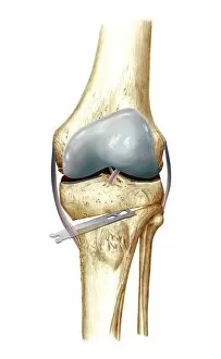
Background imageLigaments Collection: Knee osteotomy, artwork C016 / 6995

Knee osteotomy, artwork C016 / 6995
Knee osteotomy. Artwork of a bone tool being used during an osteotomy operation on the knee. An osteotomy involves the removal of bone

Background imageLigaments Collection: Plantarflexion of the foot, artwork C016 / 6799

Plantarflexion of the foot, artwork C016 / 6799
Plantarflexion of the foot. Artwork of the muscles of the foot from the side, with red arrows showing the direction of movement when flexing the foot in the direction of its lower (plantar)

Background imageLigaments Collection: Elbow and wrist extension, artwork C016 / 6791

Elbow and wrist extension, artwork C016 / 6791
Elbow and wrist extension. Artwork of the muscles of the arm from the front, with red arrows showing the direction of movement when extending the elbow and wrist joints

Background imageLigaments Collection: Wrist joint anatomy, artwork C016 / 6549

Wrist joint anatomy, artwork C016 / 6549
Wrist joint anatomy. Artwork of the bones (yellow) and ligaments (blue) of the wrist joint, in longitudinal section. The left hand has been sectioned in a palmar aspect to expose the synovial

Background imageLigaments Collection: Atlanto-occipital joint, artwork C016 / 6557

Atlanto-occipital joint, artwork C016 / 6557
Atlanto-occipital joint. Artwork of the bones (yellow) and ligaments (blue) of the atlanto-occipital joint, as seen from the side in a medial sagittal section. The rear of the spine is at right

Background imageLigaments Collection: Little and ring finger flexion, artwork C016 / 6794

Little and ring finger flexion, artwork C016 / 6794
Little and ring finger flexion. Artwork of the muscles of the forearm and hand from the front, with red arrows showing the direction of movement of the little and ring (fourth and third)

Background imageLigaments Collection: Vertebral joints of the neck, artwork C016 / 6551

Vertebral joints of the neck, artwork C016 / 6551
Vertebral joints of the neck. Artwork of the bones (yellow) and ligaments (blue) of the cervical (neck) region of the spine (vertebral column)

Background imageLigaments Collection: Atlanto-axial joint, artwork C016 / 6552

Atlanto-axial joint, artwork C016 / 6552
Atlanto-axial joint. Artwork of the bones (yellow) and ligaments (blue) of the atlanto-axial joint of the upper neck, as seen from above. The rear of the spine is at bottom

Background imageLigaments Collection: Atlanto-occipital joint, artwork C016 / 6553

Atlanto-occipital joint, artwork C016 / 6553
Atlanto-occipital joint. Artwork of the bones (yellow) and ligaments (blue) of the atlanto-occipital joint, as seen from behind (posterior view)

Background imageLigaments Collection: Runners anatomy, artwork

Runners anatomy, artwork
Runners anatomy. Artwork showing the musculature of a running human figure. The main muscles used when running are the quadriceps femoris (rectus femoris, vastus medialis, vastus lateralis)

Background imageLigaments Collection: Knee bones and ligaments, artwork C016 / 7011

Knee bones and ligaments, artwork C016 / 7011
Knee bones and ligaments. Artwork of a frontal view of the knee joint, showing bones, ligaments, tendons and cartilage. The bones are the femur (thigh bone, top), patella (knee-cap, upper centre)

Background imageLigaments Collection: Atlanto-occipital joint, artwork C016 / 6556

Atlanto-occipital joint, artwork C016 / 6556
Atlanto-occipital joint. Artwork of the bones (yellow) and ligaments (blue) of the atlanto-occipital joint, as seen from behind (posterior view)

Background imageLigaments Collection: Elbow and wrist extension, artwork C016 / 6792

Elbow and wrist extension, artwork C016 / 6792
Elbow and wrist extension. Artwork of the muscles of the arm from the front, with red arrows showing the direction of movement when extending the elbow and wrist joints


All Professionally Made to Order for Quick Shipping

"Ligaments: The Unsung Heroes of Our Body's Stability and Mobility" Damaged knee ligament, artwork: A visual representation of the intricate network in our knees, highlighting the importance of their role in supporting joint stability. Skeleton & Ligaments: Explore the hidden framework beneath our skin as we delve into the fascinating connection between our skeleton and ligaments that enable us to move with grace. Arm circulation, anatomical artwork C013 / 7419: Discover how ligaments work hand-in-hand with blood vessels to ensure proper arm circulation, allowing us to perform everyday tasks effortlessly. Muscles of the neck: Unveil the interplay between muscles and ligaments in our neck region, crucial for maintaining posture and facilitating smooth head movements. Eye muscle, TEM C014 / 1468: Delicate yet powerful eye muscles intricately intertwined with resilient ligaments—uncover this captivating combination responsible for precise eye movements. Outer ankle ligaments, artwork C013 / 4452: Step into a world where outer ankle ligaments provide stability during every stride we take—a testament to their resilience against external forces. Inner ankle ligaments, artwork C013 / 4451: Journey deeper into understanding inner ankle ligament anatomy as they safeguard against inward rolling motions while walking or running. Eye anatomy, SEM: Peer through a scanning electron microscope lens at detailed images revealing how tiny but mighty eye structures rely on robustly connected networks of supportive ligaments. Muscles (engraving): Witness an enchanting engraving showcasing various muscles working harmoniously alongside essential connective tissues like tendons and—of course—ligaments. Ligaments of the human body (engraving): An artistic masterpiece capturing an array of interconnected human body parts held together by strong bands known as "ligaments"—a true testament to the body's architectural brilliance.