Fraud Blocker Skip to main content

Kidneys Collection

"Exploring the intricate world of kidneys: a journey through art and science" Embarking on a full body scan

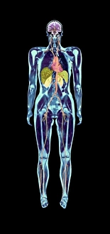
Background imageKidneys Collection: Full body scan, MRI scan

Full body scan, MRI scan
Full body scan of a female swimmer. Her brain is purple (top), her heart and arteries are red/orange (upper centre), her kidneys are pale green (centre)

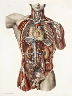
Background imageKidneys Collection: Cardiovascular system, historical artwork

Cardiovascular system, historical artwork. 19th Century hand coloured lithographic print showing the blood vessels (red and blue) of the human neck and torso

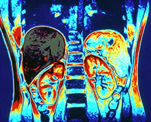
Background imageKidneys Collection: Colour MRI scan of abdomen showing kidneys & liver

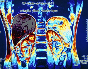
Colour MRI scan of abdomen showing kidneys & liver
Healthy kidneys. Coloured Magnetic Resonance Imaging (MRI) scan of a coronal section through a human abdomen, showing healthy kidneys and liver. A front view is seen

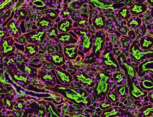
Background imageKidneys Collection: Kidney tubules in section

Kidney tubules in section
Kidney tubules. Fluorescent light micrograph of a section through kidney tissue showing numerous tubules (black/green). The tubules are seen in cross-section

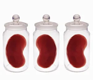
Background imageKidneys Collection: Spare kidneys

Spare kidneys. Conceptual computer artwork of spare kidneys stored in jars. This may represent the growth of human tissue to produce material for transplant surgery to replace defective or old body

Background imageKidneys Collection: Pig anatomy, artwork

Pig anatomy, artwork
Pig anatomy. Artwork showing the internal and external anatomy of the domesticated pig (Sus scrofa domestica), which is reared on farms worldwide for its meat
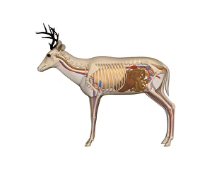
Background imageKidneys Collection: Deer anatomy, artwork

Deer anatomy, artwork
Deer anatomy. Computer artwork showing the internal anatomy of a deer (family Cervidae). Anatomical features shown here include the skeleton, lungs, intestines, bladder

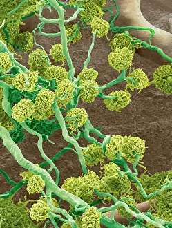
Background imageKidneys Collection: Kidney glomeruli, SEM

Kidney glomeruli, SEM
Kidney glomeruli, coloured scanning electron micrograph (SEM). The cells that usually surround the glomeruli have been removed to reveal the coiled capillaries at the core of the glomeruli

Background imageKidneys Collection: False-colour SEM showing two renal glomeruli

False-colour SEM showing two renal glomeruli
False-colour scanning electron micrograph of a section of kidney cortex, showing two glomeruli (red & green). A glomerulus is a tiny knot of blood capillaries which lies cupped within a Bowmans

Background imageKidneys Collection: Human venous system

Human venous system
LeonardoÔé¼Ôäós anatomical drawing of a man to show the heart lungs and main arteries. Drawing of a " venous figure" by Leonardo da Vinci, taken from his Anatomical note- books

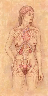
Background imageKidneys Collection: Front view of the female anatomy hilighting the endocrine system

Front view of the female anatomy hilighting the endocrine system
Brain, Anatomy, Diagram, Heart, Illustration, Bone, Shoulder, Medical, Uterus, Biology, Pelvis, Femur, See Through, Vertical, Artwork, Female Likeness, Color Image, Front View

Background imageKidneys Collection: William Bowman (1816- 1892) English anatomist, surgeon and ophthalmologist, born at Nantwich

William Bowman (1816- 1892) English anatomist, surgeon and ophthalmologist, born at Nantwich, Cheshire
542173 William Bowman (1816- 1892) English anatomist, surgeon and ophthalmologist, born at Nantwich, Cheshire. Cartoon in the Fancy Portraits series from Punch

Background imageKidneys Collection: The different varieties of Liverworts and Geranium

The different varieties of Liverworts and Geranium
5308108 The different varieties of Liverworts and Geranium.; (add.info.: Engraving depicting the different varieties of Liverworts (1,2,3) and Geranium

Background imageKidneys Collection: Advertisement for Holloway's Pills

Advertisement for Holloway's Pills
5310159 Advertisement for Holloway's Pills; (add.info.: Advertisement for Holloway's Pills, which claimed to purify blood and correct disorders of the liver, stomach, kidneys, and bowels)

Background imageKidneys Collection: Woman in costume made from betting stubs, les tickets, at the horse races, with skirt, pantaloons

Woman in costume made from betting stubs, les tickets, at the horse races, with skirt, pantaloons
FLO4684499 Woman in costume made from betting stubs, les tickets, at the horse races, with skirt, pantaloons, headdress and reins decorated with betting stubs

Background imageKidneys Collection: Anatomy of the human venal system in the upper torso. Lithograph by C

Anatomy of the human venal system in the upper torso. Lithograph by C. Mayer from Lorenz Oken's Universal Natural
FLO4684833 Anatomy of the human venal system in the upper torso. Lithograph by C. Mayer from Lorenz Oken's Universal Natural History, Allgemeine Naturgeschichte fur alle Stande, Stuttgart

Background imageKidneys Collection: Starvation, degrees of wasting (litho)

Starvation, degrees of wasting (litho)
5998374 Starvation, degrees of wasting (litho) by English School, (20th century); Private Collection; (add.info.: Starvation, degrees of wasting)

Background imageKidneys Collection: Butchers Meat (chromolitho)

Butchers Meat (chromolitho)
3651693 Butchers Meat (chromolitho) by English School, (19th century); Private Collection; (add.info.: Butchers Meat. Illustration for The Royal Cookery Book (Le Livre De Cuisine)

Background imageKidneys Collection: Diseases of the Stomach and Kidneys (colour litho)

Diseases of the Stomach and Kidneys (colour litho)
6006706 Diseases of the Stomach and Kidneys (colour litho) by European School, (19th century); Private Collection; (add.info.: Diseases of the Stomach and Kidneys)

Background imageKidneys Collection: Internal organs of the body, plate from Anatomy of the Visceras dissected

Internal organs of the body, plate from Anatomy of the Visceras dissected
CHT219321 Internal organs of the body, plate from Anatomy of the Visceras dissected, painted and engraved by Gautier, 1745 (colour engraving) by Gautier d Agoty

Background imageKidneys Collection: Anatomy of the human venal system in the upper torso

Anatomy of the human venal system in the upper torso. Lithograph by C. Mayer from Lorenz Okens Universal Natural History, Allgemeine Naturgeschichte fur alle Stande, Stuttgart, 1839

Background imageKidneys Collection: Man, his Organs, his Muscles and his Skeleton, from Anatomie Elementaire by Dr

Man, his Organs, his Muscles and his Skeleton, from Anatomie Elementaire by Dr
CHT235031 Man, his Organs, his Muscles and his Skeleton, from Anatomie Elementaire by Dr. Lelievre (colour litho) by French School, (19th century); Private Collection; (add.info.: l homme)

Background imageKidneys Collection: Anatomy, the abdominal viscera, kidneys, etc, the oestrus equi, gadfly, ova, larvae

Anatomy, the abdominal viscera, kidneys, etc, the oestrus equi, gadfly, ova, larvae, bots etc (litho)
5202581 Anatomy, the abdominal viscera, kidneys, etc, the oestrus equi, gadfly, ova, larvae, bots etc (litho) by Herring, Benjamin (1830-71); Private Collection; (add.info.: Anatomy)

Background imageKidneys Collection: Calculs des rains, Mecanisme de la colique nephretique (colour litho)

Calculs des rains, Mecanisme de la colique nephretique (colour litho)
5210391 Calculs des rains, Mecanisme de la colique nephretique (colour litho) by French School, (19th century); Private Collection; (add.info.: Calculs des rains, Mecanisme de la colique nephretique)

Background imageKidneys Collection: Anatomical View of the Human Torso, 1914 (engraving)

Anatomical View of the Human Torso, 1914 (engraving)
2622095 Anatomical View of the Human Torso, 1914 (engraving) by German School, (20th century); Private Collection; (add.info.: Vintage anatomical study of the human torso, posterior view)

Background imageKidneys Collection: Study of the Human Figure, Anterior View, Final Stage of Dissection

Study of the Human Figure, Anterior View, Final Stage of Dissection
XYC258608 Study of the Human Figure, Anterior View, Final Stage of Dissection, from A Comparative Anatomical Exposition of the Structure of the Human Body with that of a Tiger and a Common Fowl

Background imageKidneys Collection: Internal organs of a woman, plate from Recherches Anatomiques, Physiologiques

Internal organs of a woman, plate from Recherches Anatomiques, Physiologiques
CHT235024 Internal organs of a woman, plate from Recherches Anatomiques, Physiologiques, et Pathologiques sur le System Veineux by Gilbert Breschet (1784-1845) engraved by Engelmann, Paris

Background imageKidneys Collection: Farming and Sport in Newfoundland (litho)

Farming and Sport in Newfoundland (litho)
1051467 Farming and Sport in Newfoundland (litho) by Dadd, Frank (1851-1929) (after); Private Collection; (add.info.: Farming and Sport in Newfoundland)

Background imageKidneys Collection: Anatomy of human internal organs

Anatomy of human internal organs.. Anatomy of human internal organs from the front showing heart, stomach, intestines, kidneys and bladder

Background imageKidneys Collection: Woman standing, body facing forward head turned to one side, illustration overlay showing skeleton

Woman standing, body facing forward head turned to one side, illustration overlay showing skeleton and inner organs

Background imageKidneys Collection: Female anatomy, historical model

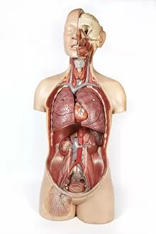
Female anatomy, historical model
Female anatomy. 1920s anatomical model showing the internal organs of the female upper body with the intestines, stomach and liver removed

Background imageKidneys Collection: Female abdominal anatomy, artwork

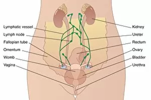
Female abdominal anatomy, artwork
Female abdominal anatomy. Computer artwork showing the kidneys and female reproductive system. Part of the lymphatic system (green) is also shown

Background imageKidneys Collection: Fig I II Caspari Bauhini Basileensis Theatrvm

Fig I II Caspari Bauhini Basileensis Theatrvm
Fig. I. Fig. II. Caspari Bauhini Basileensis Theatrvm anatomicum: novis figuris oeneis illustratum et in lucem emissum opera and sumptibus Theodori de Bry p. m

Background imageKidneys Collection: Vagus nerve anatomy, artwork C018 / 0300

Vagus nerve anatomy, artwork C018 / 0300
Vagus nerve anatomy. Artwork of a human body from the side, showing the internal anatomy and the location of the vagus nerve (yellow)

Background imageKidneys Collection: Fight-or-flight response, artwork

Fight-or-flight response, artwork
Fight-or-flight response. Artwork showing the bodys fight-or-flight response to a threat. Visual perception (yellow arrow) can trigger fear memories controlled by the brains amygdala (yellow)

Background imageKidneys Collection: Male urinary system, artwork

Male urinary system, artwork
Male urinary system. Computer artwork of a male abdomen highlighting the urinary system. The two kidneys are at upper centre

Background imageKidneys Collection: MRI scan of human abdomen showing kidneys & liver

MRI scan of human abdomen showing kidneys & liver
Abdomen. Coloured Magnetic Resonance Imaging (MRI) scan showing a coronal section through a human abdomen, front view. The lower spinal vertebrae are seen running from top to bottom through

Background imageKidneys Collection: Internal structure of human kidneys, with inset showing medulla

Internal structure of human kidneys, with inset showing medulla

Background imageKidneys Collection: Picture No. 10876992

Picture No. 10876992
Scanning electron micrograph (SEM) of Kidney glomerulus Date:

Background imageKidneys Collection: William Bowman (1816- 1892)English anatomist, surgeon and ophthalmologist was born at Nantwich

William Bowman (1816- 1892)English anatomist, surgeon and ophthalmologist was born at Nantwich, Cheshire. After five years at Birmingham General Hospital, in 1837 he moved to Kings College Hospital

Background imageKidneys Collection: William Bowman (1816- 1892) English anatomist, surgeon and ophthalmologist was born at Nantwich

William Bowman (1816- 1892) English anatomist, surgeon and ophthalmologist was born at Nantwich, Cheshire. After five years at Birmingham General Hospital, in 1837 he moved to Kings College Hospital

Background imageKidneys Collection: Richard Bright (1789-1858) English physician, born at Bristol. He described the condition

Richard Bright (1789-1858) English physician, born at Bristol. He described the condition of the kidneys known as Brights disease. Engraving c1830

Background imageKidneys Collection: Selection of offal meats, close up

Selection of offal meats, close up

Background imageKidneys Collection: Abdominal aorta, 1825 artwork

Abdominal aorta, 1825 artwork
Abdominal aorta. Dissection, with some organs removed, showing the abdominal portion of the aorta (red), the main body artery

Background imageKidneys Collection: Deep abdominal organs, 1880 artwork C017 / 6913

Deep abdominal organs, 1880 artwork C017 / 6913
Deep abdominal organs. Historical anatomical artwork of a dissected abdomen seen from the front, showing the deep-seated organs and major blood vessels after the removal of the intestines


All Professionally Made to Order for Quick Shipping

"Exploring the intricate world of kidneys: a journey through art and science" Embarking on a full body scan, we delve into the depths of our anatomy to uncover the secrets held within. The MRI scan reveals the complex network of our cardiovascular system, intertwining with historical artwork that depicts its significance throughout time. Zooming in further, we encounter kidney tubules in section - tiny structures responsible for filtering waste from our blood. A vivid colour MRI scan showcases the exquisite placement of these vital organs alongside the liver in our abdomen. Did you know? We actually possess spare kidneys. Just like pig anatomy depicted in ancient artwork, humans have two kidneys but can function with just one if needed. Our bodies are truly remarkable. Taking an even closer look, we witness the mesmerizing beauty of kidney glomeruli under scanning electron microscopy (SEM). These intricate structures play a crucial role in filtration and maintaining fluid balance within our bodies. As we explore deeper into our venous system, we marvel at its complexity and interconnectedness. X-ray images reveal abdominal arteries pulsating with life - nourishing every corner of our being. Returning to SEM imagery once again, we observe the delicate web of blood vessels surrounding each kidney glomerulus, and is here that filtration occurs, ensuring waste products are efficiently removed from our bloodstream. In this captivating journey through art and science, we gain a newfound appreciation for these incredible organs known as kidneys. They not only filter waste but also regulate electrolyte levels and maintain overall homeostasis within us – silently working day and night to keep us healthy.