Fraud Blocker Skip to main content

Kidneys Collection (#4)

"Exploring the intricate world of kidneys: a journey through art and science" Embarking on a full body scan

Background imageKidneys Collection: Kidneys, abdominal CT scan

Kidneys, abdominal CT scan
Normal kidneys. Coloured axial (horizontal) CT (computed tomography) scan through a human abdomen at the level of the kidneys. The front of the body is at top

Background imageKidneys Collection: Artwork of a section through the human kidney

Artwork of a section through the human kidney
Kidney. Artwork of a section through part of a human kidney. Coloured pink is the medulla. Contained within the medulla are the functional units of the kidney, the nephrons

Background imageKidneys Collection: Coloured SEM of podocytes in the human kidney

Coloured SEM of podocytes in the human kidney
Kidney glomerulus. Coloured scanning electron micrograph (SEM) of podocyte cells on a glomerulus in the kidney. Glomeruli are made up of a tight knot of blood capillaries covered in podocyte cells

Background imageKidneys Collection: Torso blood vessels

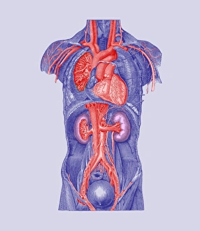
Torso blood vessels. Historical artwork of a human torso that has been dissected to show major blood vessels. The heart (upper centre) pumps blood to the lungs on either side (to be oxygenated)

Background imageKidneys Collection: Diuresis

Diuresis. Conceptual image of the effect of diuretics on the kidney. A diuretic is any drug that encourages the excretion of urine. Common diuretics include caffeine and alcohol

Background imageKidneys Collection: Diuretic effect, computer artwork

Diuretic effect, computer artwork
Diuretic effect. Computer artwork of the effect of diuretics (grey barriers) on a renal tubule (red) inside the kidney. These tubules regulate the levels of chemicals such as sodium (orange)

Background imageKidneys Collection: Pancreatitis, CT scan

Pancreatitis, CT scan
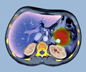
Pancreatitis. Coloured computed tomography (CT) scan of an axial section through a patients abdomen showing a false aneurysm (red) due to chronic pancreatitis. The front of the body is at top

Background imageKidneys Collection: Kidney glomeruli, light micrograph

Kidney glomeruli, light micrograph
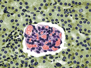
Kidney glomeruli. Light microscope of a section through the cortex of a healthy kidney showing glomeruli. The core of a glomerulus is formed of a tightly coiled capillary (purple)

Background imageKidneys Collection: Kidney capillary, SEM

Kidney capillary, SEM
Kidney capillary. Coloured scanning electron micrograph (SEM) of a fracture through a glomerular capillary. A kidney glomerulus, which filters waste products from the blood

Background imageKidneys Collection: Kidney tubule cell, coloured SEM

Kidney tubule cell, coloured SEM
Kidney tubule cell, coloured scanning electron micrograph (SEM). This cell (brown) is covered in thin projections (microvilli) that serve to increase its surface area

Background imageKidneys Collection: Coloured SEM of a renal glomerulus

Coloured SEM of a renal glomerulus
Glomerulus. Coloured Scanning Electron Micrograph (SEM) of a glomerulus - a tight knot of blood capillaries (yellow) inside one of the kidneys microscopic blood-filtering units

Background imageKidneys Collection: Computer graphic of kidneys (waterdrop background)

Computer graphic of kidneys (waterdrop background)
Kidneys. Computer graphic illustration of two human kidneys, seen on a waterdrop background to depict the role of kidneys as fluid filtration units

Background imageKidneys Collection: Artwork of kidney nephron with blood vessels

Artwork of kidney nephron with blood vessels
Nephron. Illustration of a nephron from the human kidney, surrounded by blood vessels. Nephrons are where waste products and excess water are removed from the bloodstream in the kidney

Background imageKidneys Collection: Computer graphic of section through human kidney

Computer graphic of section through human kidney
Kidney. Computer graphic of a section through a whole human kidney. At upper left is the renal artery and vein which supply blood to, and drain blood from the kidney

Background imageKidneys Collection: Illustration of nephron structure in human kidney

Illustration of nephron structure in human kidney
Illustration of a nephron, the functional unit of excretion in the human kidney. As a blood filtering system, a nephron is richly supplied with capillaries from branches of the renal artery (red)

Background imageKidneys Collection: Renal corpuscle of kidney

Renal corpuscle of kidney
Coloured scanning electron micrograph (SEM) of a renal corpuscle in the kidney. The renal corpuscle is formed by the glomerulus (red), the convoluted capillary at centre

Background imageKidneys Collection: Kidneys

Kidneys
Illustration of the human kidneys. The arterial blood supply to the kidneys is through the renal arteries (red), which branch from the abdominal aorta

Background imageKidneys Collection: Kidney infection

Kidney infection. Artwork of a section through the urinary system showing an infection of a kidney (pyelonephritis, upper left)

Background imageKidneys Collection: Human urinary system, artwork

Human urinary system, artwork
Urinary system. Computer artwork of a male torso and the urinary system, showing kidney, urethra, bladder and suprarenal gland

Background imageKidneys Collection: Healthy kidneys, artwork

Healthy kidneys, artwork

Background imageKidneys Collection: Healthy urinary system, artwork

Healthy urinary system, artwork

Background imageKidneys Collection: Kidneys, artwork

Kidneys, artwork
Kidneys, computer artwork

Background imageKidneys Collection: Human kidneys, artwork

Human kidneys, artwork
Human kidneys, computer artwork

Background imageKidneys Collection: Circulatory system, artwork

Circulatory system, artwork

Background imageKidneys Collection: Human circulatory system, artwork

Human circulatory system, artwork
Human circulatory system and red blood cells, computer artwork. Veins are blue and arteries are red

Background imageKidneys Collection: Human internal organs, diagram

Human internal organs, diagram
Human internal organs. Diagram showing the human internal organs in the head and torso. In the head, the organs and cavities shown are: the brain (encephalon), the cerebellum (part of the brain)

Background imageKidneys Collection: Circulatory system, 16th century

Circulatory system, 16th century
Circulatory system. 16th-century artwork showing the heart and the arteries supplying blood to the body, as drawn by the Belgian anatomist Andreas Vesalius (1514-1564)

Background imageKidneys Collection: Human internal organs, artwork

Human internal organs, artwork
Human internal organs. Artwork showing the human internal organs in the head and torso. In the head, the organs and cavities shown are: the brain (encephalon), the cerebellum (part of the brain)

Background imageKidneys Collection: Diuretic effect, artwork

Diuretic effect, artwork
Diuretic effect. Computer artwork showing the effect of diuretics (grey barriers) on a renal tubule (red) inside the kidney

Background imageKidneys Collection: Urinary system, artwork

Urinary system, artwork
Urinary system. Artwork showing the human urinary system on an outline of the human body, showing the location of the kidneys (upper left and upper right) and bladder (lower centre)

Background imageKidneys Collection: Calcified aortic artery, 3D CT scan

Calcified aortic artery, 3D CT scan
Coloured three-dimensional (3D) Computed Tomography (CT) scan in front view of the abdomen in a patient showing the kidneys (red)

Background imageKidneys Collection: Spine and aorta, 3D CT scan

Spine and aorta, 3D CT scan
Coloured three-dimensional (3D) Computed Tomography (CT) scan in front view of the abdomen in a patient showing the vertebral bones of the spine (running top to bottom)

Background imageKidneys Collection: Medullary sponge kidney, CT scan

Medullary sponge kidney, CT scan
Medullary sponge kidney. Coloured computed tomography (CT) scan in axial section of the abdomen of a 25-year-old patient with medullary sponge kidney, or Cacchi Ricci disease

Background imageKidneys Collection: Kidneys and bladder, 3D CT scan

Kidneys and bladder, 3D CT scan
Coloured three-dimensional (3D) Computed Tomography (CT) scan in front view of the abdomen in a patient showing the kidneys (blue) on either side of the vertebral bones of the spine (at upper centre)

Background imageKidneys Collection: Aortic aneurysm CT scan

Aortic aneurysm CT scan
Aortic aneurysm, coloured 3D computed tomography (CT) scan. Side view of the abdomen of a 65-year-old patient with an extended aneurysm of the aorta

Background imageKidneys Collection: Abdominal aortic aneurysm, artwork

Abdominal aortic aneurysm, artwork
Abdominal aortic aneurysm. Computer artwork showing a large aneurysm (round, centre) in the abdominal section of the aorta

Background imageKidneys Collection: Penguins mating, artwork

Penguins mating, artwork. The penguins reproductive organs are shown here, with the male on top and the female on the bottom

Background imageKidneys Collection: Whale anatomy

Whale anatomy. Artworks showing a dead beached whale (top) and its internal anatomy (bottom). Whales are marine mammals that swim using their flippers and tails, and surface to breathe

Background imageKidneys Collection: Artwork of the kidneys

Artwork of the kidneys, the organs involved in excretion of waste from the blood as urine through the ureters (tubes, lower frame). Renal blood vessels are seen

Background imageKidneys Collection: Melamine and kidneys, artwork

Melamine and kidneys, artwork
Melamine and kidneys, computer artwork. Melamine is an organic base with a high nitrogen content. Melamine and formaldehyde are the main components of the plastic known as melamine resin

Background imageKidneys Collection: Kidney glomerulus, light micrograph

Kidney glomerulus, light micrograph
Kidney glomerulus. Coloured light micrograph of a section through the cortex of a healthy kidney, showing a glomerulus. The core of a glomerulus is formed of a tightly coiled capillary (purple)

Background imageKidneys Collection: Female urinary system, artwork

Female urinary system, artwork
Female urinary system. Computer artwork of a naked woman with a healthy urinary system. The kidneys can be seen (upper left and right), as well as the ureters

Background imageKidneys Collection: Aortic aneurysm, 3-D CT scan

Aortic aneurysm, 3-D CT scan
3-D Computed Tomography (CT) scan of the abdomen of a patient showing an aortic aneurysm, a bulging of this main artery of the body (red, at centre) which can cause it to rupture if not treated

Background imageKidneys Collection: Womans abdomen, 17th century artwork

Womans abdomen, 17th century artwork. Dissection showing a womans abdomen opened with the intestines removed, to show the inner abdominal organs


All Professionally Made to Order for Quick Shipping

"Exploring the intricate world of kidneys: a journey through art and science" Embarking on a full body scan, we delve into the depths of our anatomy to uncover the secrets held within. The MRI scan reveals the complex network of our cardiovascular system, intertwining with historical artwork that depicts its significance throughout time. Zooming in further, we encounter kidney tubules in section - tiny structures responsible for filtering waste from our blood. A vivid colour MRI scan showcases the exquisite placement of these vital organs alongside the liver in our abdomen. Did you know? We actually possess spare kidneys. Just like pig anatomy depicted in ancient artwork, humans have two kidneys but can function with just one if needed. Our bodies are truly remarkable. Taking an even closer look, we witness the mesmerizing beauty of kidney glomeruli under scanning electron microscopy (SEM). These intricate structures play a crucial role in filtration and maintaining fluid balance within our bodies. As we explore deeper into our venous system, we marvel at its complexity and interconnectedness. X-ray images reveal abdominal arteries pulsating with life - nourishing every corner of our being. Returning to SEM imagery once again, we observe the delicate web of blood vessels surrounding each kidney glomerulus, and is here that filtration occurs, ensuring waste products are efficiently removed from our bloodstream. In this captivating journey through art and science, we gain a newfound appreciation for these incredible organs known as kidneys. They not only filter waste but also regulate electrolyte levels and maintain overall homeostasis within us – silently working day and night to keep us healthy.