Canvas Print : Microscopic Objects


![]()
Canvas Prints from Mary Evans Picture Library
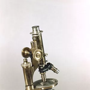
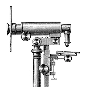
Microscopic Objects
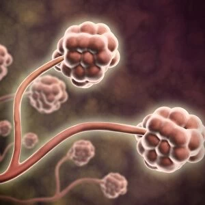
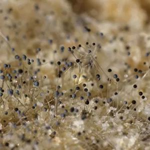
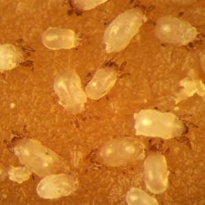
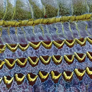
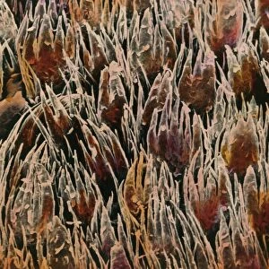
A variety of living and non- living objects magnified through a microscope
Mary Evans Picture Library makes available wonderful images created for people to enjoy over the centuries
Media ID 589701
© Mary Evans Picture Library 2015 - https://copyrighthub.org/s0/hub1/creation/maryevans/MaryEvansPictureID/10179015
1810 Antennae Cells Crystals Instruments Living Magnified Microscope Microscopic Sperm Variety Veins Follicles
20"x16" (51x41cm) Canvas Print
Discover the fascinating world of microscopic objects with Media Storehouse's Canvas Prints from Mary Evans Prints Online. These captivating prints showcase an intriguing collection of living and non-living specimens, magnified to reveal their hidden beauty. Each print is meticulously captured through a microscope, revealing intricate details and textures that are often unseen to the naked eye. These high-quality canvas prints are not only a stunning addition to any room but also serve as a conversation starter, inspiring curiosity and wonder. Bring the microscopic world into your home and elevate your decor with these unique and captivating prints.
Delivered stretched and ready to hang our premium quality canvas prints are made from a polyester/cotton blend canvas and stretched over a 1.25" (32mm) kiln dried knot free wood stretcher bar. Packaged in a plastic bag and secured to a cardboard insert for safe transit.
Canvas Prints add colour, depth and texture to any space. Professionally Stretched Canvas over a hidden Wooden Box Frame and Ready to Hang
Estimated Product Size is 40.6cm x 50.8cm (16" x 20")
These are individually made so all sizes are approximate
Artwork printed orientated as per the preview above, with portrait (vertical) orientation to match the source image.
EDITORS COMMENTS
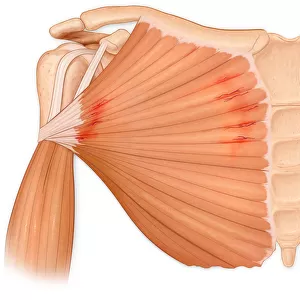
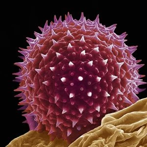
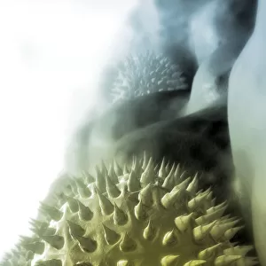
This print showcases a fascinating array of microscopic objects, providing a glimpse into the intricate world that exists beyond what the naked eye can see. Dating back to circa 1810, this historical image captures a variety of living and non-living objects that have been magnified through a microscope.
From sperm cells to crystals, veins to follicles, this collection of magnified specimens offers a unique perspective on the beauty and complexity of the natural world. Each object is meticulously detailed, revealing intricate patterns and structures that are both mesmerizing and educational.
The instruments used to capture these images are also featured in the print, adding an extra layer of historical significance to this piece. It serves as a reminder of the advancements made in science and technology over the years, allowing us to explore and understand our world in ways that were once unimaginable.
Whether you're interested in science, history, or simply appreciate the beauty of nature at its smallest scale, this print is sure to captivate your imagination. Take a closer look at these microscopic wonders and marvel at the hidden treasures waiting to be discovered within each tiny cell or crystal.
MADE IN THE USA
Safe Shipping with 30 Day Money Back Guarantee
FREE PERSONALISATION*
We are proud to offer a range of customisation features including Personalised Captions, Color Filters and Picture Zoom Tools
SECURE PAYMENTS
We happily accept a wide range of payment options so you can pay for the things you need in the way that is most convenient for you
* Options may vary by product and licensing agreement. Zoomed Pictures can be adjusted in the Cart.