Fraud Blocker Skip to main content

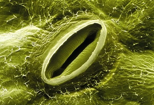
Open stoma pore on rose leaf

Open stoma pore on rose leaf
In Stock
We accept all major payment options so you can pay for the things you need in the way that is most convenient for you
Framed Photos
Buy from $79.99
Photo Prints
Buy from $9.99
Jigsaw Puzzles
Buy from $39.99
Poster Prints
Buy from $24.99
Canvas Prints
Buy from $99.99
Fine Art Prints
Buy from $60.90
Metal Prints
Buy from $169.99
Home Decor
Buy from $16.99

Science Photo Library Photo Prints and Wall Art

Open stoma pore on rose leaf

Stoma on a rose leaf. Coloured scanning electron micrograph (SEM) of the lower leaf surface of a Garden Rose Rosa sp. showing an open stoma. Epidermal cells (green) occur around this structure. A stoma (plural: stomata) is a tiny pore bordered by two kidney-shaped guard cells. Here the partly open pore is elongated in shape. Guard cells serve to open and close the stoma: opening the pore allows gases to be exchanged by leaf tissues during photosynthesis; and closing the pore at night or during dry periods prevents water loss. In the rose, more stomata occur on the underside of leaves. Magnification: x850 at 5x7cm size. Magnification: x2, 500 at 6x4.5inch size. Science Photo Library features Science and Medical images including photos and illustrations. © POWER AND SYRED/SCIENCE PHOTO LIBRARY

Media ID 9194273

Open Plants Rose Stoma Stomata

SECURE PAYMENTS
We accept all major payment options so you can pay for the things you need in the way that is most convenient for you

Made in USA
Safe Shipping, Easy Returns

FREE PERSONALISATION
We are proud to offer a range of customisation including Cropping, Captions, and Filters. Zoom Cropping can be adjusted in the basket


EDITORS COMMENTS

This print from Science Photo Library showcases the intricate beauty of nature at a microscopic level. The image captures an open stoma pore on a rose leaf, revealing the fascinating inner workings of this delicate structure. In vibrant colors, the scanning electron micrograph highlights the lower surface of a Garden Rose Rosa sp. leaf. Surrounding the stoma are epidermal cells in shades of green, emphasizing their role in protecting and supporting this vital opening. Stomata, like the one displayed here, are tiny pores bordered by two kidney-shaped guard cells. This particular stoma appears elongated in shape and is partly open to facilitate gas exchange during photosynthesis. By opening up, it allows leaf tissues to absorb essential gases while closing at night or during dry periods prevents water loss. The rose plant has strategically placed stomata primarily on its underside leaves for optimal efficiency. Through magnification at 850 times for a 5x7cm size or 2,500 times for a 6x4.5inch size, we can truly appreciate the intricate details that make up this natural wonder. This stunning photograph serves as a reminder of how even something as small as an open stoma pore can play such a crucial role in sustaining life within our botanical world.


Framed Prints of Open stoma pore on rose leaf

Discover the intricacies of nature with our Media Storehouse Framed Prints featuring the captivating "Open Stoma Pore on Rose Leaf" image. This coloured Scanning Electron Micrograph (SEM) reveals the stunning detail of a Garden Rose Rosa sp. leaf, showcasing an open stoma surrounded by epidermal cells. Bring the beauty of science into your home or office with this unique and thought-provoking addition to your décor.


Photo Prints of Open stoma pore on rose leaf

Discover the intricacies of nature with Media Storehouse's Photographic Prints. This captivating image, sourced from Science Photo Library, showcases an open stoma pore on a Garden Rose leaf. The vibrant colored scanning electron micrograph (SEM) reveals the intricate structure of the lower leaf surface, with epidermal cells surrounding the stoma. Bring this breathtaking display of nature into your home or office and ignite curiosity with every glance.


Poster Prints of Open stoma pore on rose leaf

Discover the beauty of nature's intricacies with our Media Storehouse Poster Prints featuring the captivating image of an open stoma on a rose leaf. This coloured Scanning Electron Micrograph (SEM) reveals the intricate details of the Garden Rose Rosa sp.'s epidermal cells surrounding the stoma, creating a stunning visual representation of the plant's respiratory system. Bring this scientific marvel into your home or office, and ignite curiosity and inspiration with every glance.


Jigsaw Puzzles of Open stoma pore on rose leaf

Discover the intricacies of nature with Media Storehouse's Science-themed Jigsaw Puzzles. This captivating puzzle features an Open Stoma Pore on a Rose Leaf, captured in stunning detail through Science Photo Library's Coloured Scanning Electron Micrograph (SEM). Delve into the microscopic world of a Garden Rose (Rosa sp.) and piece together the intricate patterns of epidermal cells surrounding the open stoma. Engage your mind, challenge your dexterity, and deepen your appreciation for the beauty of science with this unique and educational jigsaw puzzle.

diamond
Made With High Grade Materials
thumb_up
Professional Quality Finish
package_2
Carefully Packed To Aid Safe Arrival

You May Also Like


Dry-stone walls and barns in Wensleydale, Yorkshire Dales National Park, North YorkshireDry-stone walls and barns in Wensleydale, Yorkshire Dales National Park, North Yorkshire
Add to Basket
Pacific Islands, Oceania: group at a celebrationPacific Islands, Oceania: group at a celebration
Add to Basket
Brassy chub, Kyphosus vaigiensisBrassy chub, Kyphosus vaigiensis
Add to Basket
Romantic paper lace card - Remember MeRomantic paper lace card - Remember Me
Add to Basket
Westland Lysander R9125Westland Lysander R9125
Add to Basket
Canterbury Tales - The KnightCanterbury Tales - The Knight
Add to Basket
Broadwoods Pianoforte manufactory 1858Broadwoods Pianoforte manufactory 1858
Add to Basket
Santiago de Compostela, Spain - Palacio ConsistorialSantiago de Compostela, Spain - Palacio Consistorial
Add to Basket
Macrophage, SEMMacrophage, SEM
Add to Basket
Three Wild Boar (Sus scrofa) foraging in snow. The Netherlands, JanuaryThree Wild Boar (Sus scrofa) foraging in snow. The Netherlands, January
Add to Basket
Giambattista BodoniGiambattista Bodoni
Add to Basket
The Standard of England - Girl Guides AssociationThe Standard of England - Girl Guides Association
Add to Basket

Gallery Wall Inspiration

Nerve cell, SEM
False-coloured SEM (Scanning Electron Micrograph) of a Tardigrade (Tardigrada) amongst moss, from Peak District National Park, Derbyshire, UK. This tardigrade is approximately 0. 1 millimetres long. Digital composite
Santos-Dumont / Sem
Neutrophil engulfing MRSA, SEM C018 / 8596
X and Y chromosomes
Lavender pollen grain, SEM
Scanning Electron Micrograph (SEM): Tardigrade or ‘Water Bear Magnification x 1250 (A4 size: 29. 7 cm width)
Cat tongue surface, SEM
Hazel pollen grains
E. coli bacteria, SEM
Collagen, Scanning electron micrograph (SEM)
Scanning electron micrograph (SEM) of red blood cell
T lymphocytes and cancer cell, SEM C001 / 1679
Tardigrade or Water Bear
Crysotile asbestos
Uterus lining during menstruation, SEM
Iris of the eye, SEM
Stylus in LP record
Macrophage engulfing TB bacteria, SEM
Salmonella bacteria, SEM
SEM of penicillin fungus
Vascular bundle, SEM
Geranium anther, SEM
Dahlia flower pollen, SEM
Scanning Electron Micrograph (SEM): Praying Mantis - Magnification x 30 (if print A4 size: 29. 7 cm wide)
Tardigrade, SEM
Botanik Digitalis purpurea L. Fingerhut 160: 1


© Copyright Media Storehouse, All Rights Reserved 2003 - 2024
Facebook     Pinterest     100% Payment Secure     Reviews IO