Home > Popular Themes > Human Body
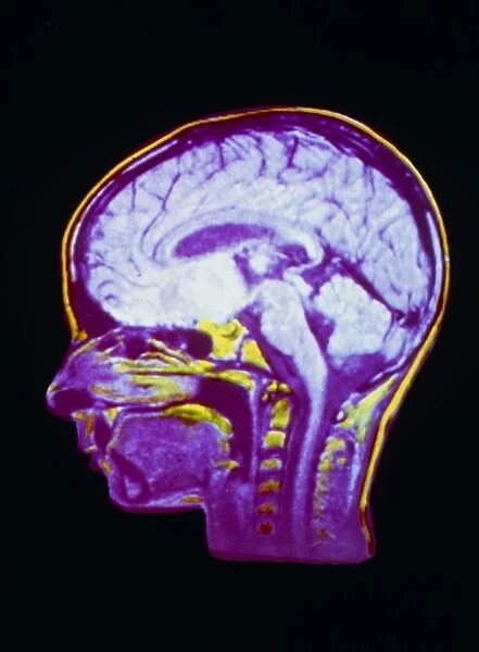
NMR scan of a childs head, sagittal section
![]()

Wall Art and Photo Gifts from Science Photo Library
NMR scan of a childs head, sagittal section
Nuclear Magnetic Resonance (NMR) image of a sagittal (mid-profile) section through a childs head, showing structures of the brain and spinal cord. Details of the brain visible include the folds of the cerebral cortex, the cerebellum (below the rear lobes of the brain hemispheres) and the pons and medulla of the brainstem, the areas of swelling at the top of the spinal cord, which can be seen running down the neck. Unlike X- ray (CT) scanning, NMR imaging does not use ionising radiations; an image is obtained by studying the radio-response of protons in body tissues that are subjected to a strong, pulsed magnetic field
Science Photo Library features Science and Medical images including photos and illustrations
Media ID 6421606
© CNRI/SCIENCE PHOTO LIBRARY
EDITORS COMMENTS
This print showcases a remarkable NMR scan of a child's head, specifically capturing a sagittal section. The image provides an intricate view of the brain and spinal cord structures, offering valuable insights into the complexities of our nervous system. Upon closer examination, one can observe the intricately folded cerebral cortex, which plays a crucial role in various cognitive functions. Positioned below the rear lobes of the brain hemispheres lies the cerebellum, responsible for coordinating movement and balance. Additionally, this NMR scan reveals the pons and medulla of the brainstem - vital regions involved in controlling essential bodily functions. Notably different from X-ray (CT) scanning methods that employ ionizing radiation, NMR imaging utilizes strong pulsed magnetic fields to study radio-responses from protons within body tissues. This non-invasive technique allows for detailed visualization without any harmful effects on health. Furthermore, this awe-inspiring photograph captures areas of swelling at the top of the spinal cord as it extends down through the neck region. Such comprehensive imaging aids medical professionals in diagnosing conditions related to both neurological development and potential injuries affecting these critical regions. Science Photo Library presents this extraordinary piece as part of their collection focused on human anatomy and neuroscience research. It serves as a testament to how advanced technology like NMR scans continues to deepen our understanding of human physiology while prioritizing patient safety above all else.
MADE IN THE USA
Safe Shipping with 30 Day Money Back Guarantee
FREE PERSONALISATION*
We are proud to offer a range of customisation features including Personalised Captions, Color Filters and Picture Zoom Tools
SECURE PAYMENTS
We happily accept a wide range of payment options so you can pay for the things you need in the way that is most convenient for you
* Options may vary by product and licensing agreement. Zoomed Pictures can be adjusted in the Cart.

