Myenteric nerve plexus, TEM

![]()
Wall Art and Photo Gifts from Science Photo Library
Myenteric nerve plexus, TEM
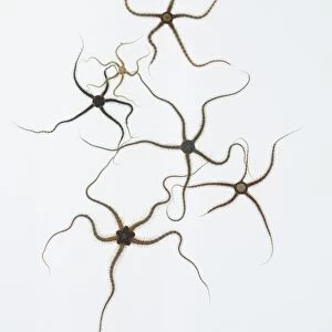
Myenteric nerve plexus. Transmission electron micrograph (TEM) of a section through a bundle of unmyelinated nerve fibres in the myenteric nerve plexus of the intestinal tract. The myenteric, or Auerbach s, plexus is a vast network of intrinsic nerve fibres and their cell bodies (ganglia) located between the layers of smooth muscle in the wall of the gut. It is part of the enteric nervous system (ENS). The events controlled, at least in part, by the ENS include: motor activity, secretion, absorption, blood flow, and interaction with other organs such as the gallbladder or pancreas. Although the ENS functions autonomously it is modified by an external nerve supply. Magnification: x6, 000 when printed 10 centimetres wide
Science Photo Library features Science and Medical images including photos and illustrations
Media ID 9242225
© MICROSCAPE/SCIENCE PHOTO LIBRARY
Black And White Bodies Bowel Bowels Cell Biology Cell Body Collection Cytological Cytology Digestive System Enteric Fibre Fibres Gastrointestinal Tract Histological Histology Intestinal Intestine Intestines Nerve Nerve Fibre Nerves Neuron Neurone Neurones Neurons Organelle Organelles Smooth Muscle Transmission Electron Micrograph Transmission Electron Microscope Unmyelinated Wall Cells Nervous System Neurological Neurology Section Sectioned
EDITORS COMMENTS
This print showcases the intricate beauty of the myenteric nerve plexus, captured using a transmission electron microscope (TEM). The image reveals a section through a bundle of unmyelinated nerve fibers within the myenteric plexus, which is located between the layers of smooth muscle in the intestinal tract. The myenteric plexus forms an extensive network of intrinsic nerve fibers and ganglia that play a crucial role in regulating various functions of the enteric nervous system (ENS). This autonomous system controls essential processes such as motor activity, secretion, absorption, blood flow, and interactions with neighboring organs like the gallbladder or pancreas. Although it operates independently, external nerves can modify its functioning. At a magnification of x6,000 when printed 10 centimeters wide, this photograph provides an up-close view into the microscopic world of cell biology and neurology. It highlights not only the structural complexity but also emphasizes how these cells work together to ensure proper digestive function. With its monochrome aesthetic and detailed depiction of organelles within neurons and their bodies known as ganglia, this image serves as both an artistic representation and scientific tool for studying histology. It offers valuable insights into our understanding of gastrointestinal health by shedding light on key components involved in maintaining bowel function.
MADE IN THE USA
Safe Shipping with 30 Day Money Back Guarantee
FREE PERSONALISATION*
We are proud to offer a range of customisation features including Personalised Captions, Color Filters and Picture Zoom Tools
SECURE PAYMENTS
We happily accept a wide range of payment options so you can pay for the things you need in the way that is most convenient for you
* Options may vary by product and licensing agreement. Zoomed Pictures can be adjusted in the Cart.