Fraud Blocker Skip to main content

Home > Science Photo Library > Images Dated > 2013 > May > 7 May 2013

Images Dated 7th May 2013

Choose a picture from our Images Dated 7th May 2013 Collection for your Wall Art and Photo Gifts

40 items

We are proud to offer this selection in partnership with Science Photo Library

Background imageImages Dated 7th May 2013: Trachea lining, SEM C015 / 9936

Trachea lining, SEM C015 / 9936
Trachea lining. Coloured scanning electron micrograph (SEM) of a section through the lining of the trachea (wind pipe). The trachea links the larynx (voice box) to the lungs

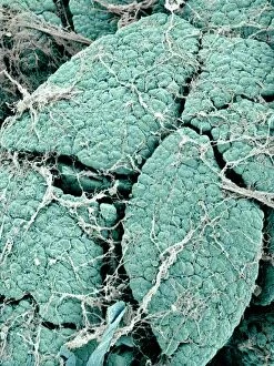
Background imageImages Dated 7th May 2013: Pancreas lobes, SEM C015 / 9499

Pancreas lobes, SEM C015 / 9499
Pancreas lobes, coloured scanning electron micrograph (SEM). Several lobes of the pancreas are seen here, separated by fissures. The smaller sections seen on each lobe are clusters of acini cells

Background imageImages Dated 7th May 2013: Liver, SEM C015 / 9924

Liver, SEM C015 / 9924
Liver vein. Coloured scanning electron micrograph (SEM) of liver tissue. The functions of the liver include destroying old red blood cells

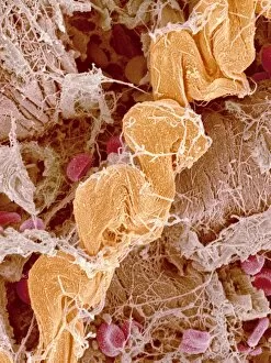
Background imageImages Dated 7th May 2013: Heart tissue, SEM C015 / 9598

Heart tissue, SEM C015 / 9598
Heart tissue. Coloured scanning electron micrograph (SEM) of heart tissue with red blood cells (erythrocytes, red) and connective tissue (orange)

Background imageImages Dated 7th May 2013: Pancreas cells, SEM C015 / 9502

Pancreas cells, SEM C015 / 9502
Pancreas cells, coloured scanning electron micrograph (SEM). The small sections seen here are clusters of acini cells. These are exocrine cells, secreting digestive enzymes

Background imageImages Dated 7th May 2013: Trachea lining, SEM C015 / 9939

Trachea lining, SEM C015 / 9939
Trachea lining. Coloured scanning electron micrograph (SEM) of a section through the lining of the trachea (wind pipe). The trachea links the larynx (voice box) to the lungs

Background imageImages Dated 7th May 2013: Pancreas lobes, SEM C015 / 9496

Pancreas lobes, SEM C015 / 9496
Pancreas lobes, coloured scanning electron micrograph (SEM). Several lobes of the pancreas are seen here, separated by fissures. The smaller sections seen on each lobe are clusters of acini cells

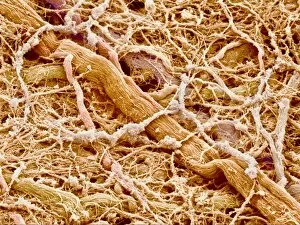
Background imageImages Dated 7th May 2013: Connective tissue, SEM C015 / 9929

Connective tissue, SEM C015 / 9929
Connective tissue. Coloured scanning electron micrograph (SEM) of connective tissue from the trachea (wind pipe). Connective tissue supports other tissues and organs

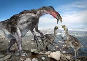
Background imageImages Dated 7th May 2013: Nothronychus dinosaur family, artwork C015 / 9714

Nothronychus dinosaur family, artwork C015 / 9714
Nothronychus dinosaur family, artwork. This theropod dinosaur, found in what is now North America some 91 million years ago, lived during the Cretaceous. The type specimen is Nothronychus mckinleyi

Background imageImages Dated 7th May 2013: Trachea lining, SEM C015 / 9610

Trachea lining, SEM C015 / 9610
Trachea lining. Coloured scanning electron micrograph (SEM) of the lining of the trachea (wind pipe). The trachea links the larynx to the lungs

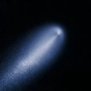
Background imageImages Dated 7th May 2013: Comet ISON, April 2013 C015 / 9817

Comet ISON, April 2013 C015 / 9817
Comet ISON (C/2012 S1), as observed on 10 April 2013 by the Hubble Space Telescope (HST). This comet was discovered on 21 September 2012 by the International Scientific Optical Network (ISON)

Background imageImages Dated 7th May 2013: Pancreas lobes, SEM C015 / 9498

Pancreas lobes, SEM C015 / 9498
Pancreas lobes, coloured scanning electron micrograph (SEM). Several lobes of the pancreas are seen here, separated by fissures. The smaller sections seen on each lobe are clusters of acini cells

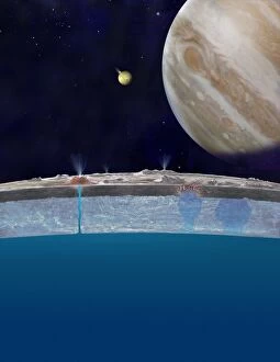
Background imageImages Dated 7th May 2013: Europas ocean, artwork

Europas ocean, artwork
Europas ocean. Artwork based on astronomical research studying the frozen surface of Europa, one of the four largest moons of Jupiter

Background imageImages Dated 7th May 2013: Connective tissue, SEM C015 / 9933

Connective tissue, SEM C015 / 9933
Connective tissue. Coloured scanning electron micrograph (SEM) of connective tissue from the trachea (wind pipe). Connective tissue supports other tissues and organs

Background imageImages Dated 7th May 2013: Pancreas cells, SEM C015 / 9501

Pancreas cells, SEM C015 / 9501
Pancreas cells, coloured scanning electron micrograph (SEM). The small sections seen here are clusters of acini cells. These are exocrine cells, secreting digestive enzymes

Background imageImages Dated 7th May 2013: Connective tissue, SEM C015 / 9926

Connective tissue, SEM C015 / 9926
Connective tissue. Coloured scanning electron micrograph (SEM) of connective tissue from the trachea (wind pipe). Connective tissue supports other tissues and organs

Background imageImages Dated 7th May 2013: Liver, SEM C015 / 9922

Liver, SEM C015 / 9922
Liver vein. Coloured scanning electron micrograph (SEM) of liver tissue showing a large vein (pink, up centre). The functions of the liver include destroying old red blood cells

Background imageImages Dated 7th May 2013: Pancreas lobes, SEM C015 / 9500

Pancreas lobes, SEM C015 / 9500
Pancreas lobes, coloured scanning electron micrograph (SEM). Several lobes of the pancreas are seen here, separated by fissures. The smaller sections seen on each lobe are clusters of acini cells

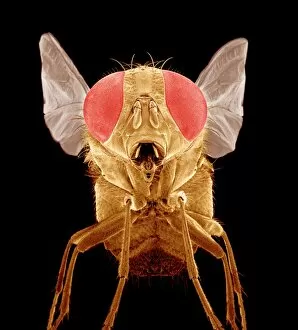
Background imageImages Dated 7th May 2013: Fly, SEM C015 / 9945

Fly, SEM C015 / 9945
Fly. Coloured scanning electron micrograph (SEM) of a fly (order Diptera). The head is dominated by two large compound eyes (red)

Background imageImages Dated 7th May 2013: Connective tissue, SEM C015 / 9928

Connective tissue, SEM C015 / 9928
Connective tissue. Coloured scanning electron micrograph (SEM) of connective tissue from the trachea (wind pipe). Connective tissue supports other tissues and organs

Background imageImages Dated 7th May 2013: Connective tissue, SEM C015 / 9932

Connective tissue, SEM C015 / 9932
Connective tissue. Coloured scanning electron micrograph (SEM) of connective tissue from the trachea (wind pipe). Connective tissue supports other tissues and organs

Background imageImages Dated 7th May 2013: Moth antenna, SEM C015 / 9946

Moth antenna, SEM C015 / 9946
Moth antenna. Coloured scanning electron micrograph (SEM) of a moths antenna, showing many sensory hairs. These hairs are used to detect touch and vibrations

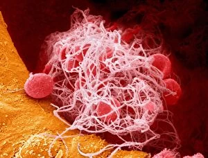
Background imageImages Dated 7th May 2013: Blood clot, SEM C015 / 9608

Blood clot, SEM C015 / 9608
Blood clot. Coloured scanning electron micrograph (SEM) of a blood clot from the inner wall of the left ventricle of a heart

Background imageImages Dated 7th May 2013: Heart tissue, SEM C015 / 9541

Heart tissue, SEM C015 / 9541
Heart tissue. Coloured scanning electron micrograph (SEM) of heart tissue with red blood cells (erythrocytes)

Background imageImages Dated 7th May 2013: Closed vocal cords, SEM C015 / 9505

Closed vocal cords, SEM C015 / 9505
Vocal cords. Coloured scanning electron micrograph of the closed vocal cords of a rat. These cords are located in the larynx, part of the neck

Background imageImages Dated 7th May 2013: Liver, SEM C015 / 9923

Liver, SEM C015 / 9923
Liver vein. Coloured scanning electron micrograph (SEM) of liver tissue. The functions of the liver include destroying old red blood cells

Background imageImages Dated 7th May 2013: Connective tissue, SEM C015 / 9930

Connective tissue, SEM C015 / 9930
Connective tissue. Coloured scanning electron micrograph (SEM) of connective tissue from the trachea (wind pipe). Connective tissue supports other tissues and organs

Background imageImages Dated 7th May 2013: Pancreas lobes, SEM C015 / 9495

Pancreas lobes, SEM C015 / 9495
Pancreas lobes, coloured scanning electron micrograph (SEM). Several lobes of the pancreas are seen here, separated by fissures. The smaller sections seen on each lobe are clusters of acini cells

Background imageImages Dated 7th May 2013: Haemoglobin, molecular model C015 / 9391

Haemoglobin, molecular model C015 / 9391
Haemoglobin, molecular model. This is deoxyhaemoglobin, the molecule in its non-oxygen bound state. Haemoglobin transports oxygen around the body in red blood cells

Background imageImages Dated 7th May 2013: Vitamin B12 import proteins C015 / 9942

Vitamin B12 import proteins C015 / 9942
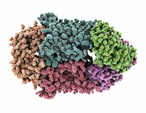
Vitamin B12 import proteins, molecular model. This complex is the import proteins btuC, btuD, and btuF. The first two together form BtuCD

Background imageImages Dated 7th May 2013: Connective tissue, SEM C015 / 9934

Connective tissue, SEM C015 / 9934
Connective tissue. Coloured scanning electron micrograph (SEM) of connective tissue from the trachea (wind pipe). Connective tissue supports other tissues and organs

Background imageImages Dated 7th May 2013: Insulin receptor molecule C015 / 9405

Insulin receptor molecule C015 / 9405
Insulin receptor, molecular model. The insulin receptor is a transmembrane protein, that is it spans the cellular membrane

Background imageImages Dated 7th May 2013: Pancreas cells, SEM C015 / 9504

Pancreas cells, SEM C015 / 9504
Pancreas cells, coloured scanning electron micrograph (SEM). The small sections seen here are clusters of acini cells. These are exocrine cells, secreting digestive enzymes

Background imageImages Dated 7th May 2013: Maggot waste digestion food production C015 / 9938

Maggot waste digestion food production C015 / 9938
Maggot waste digestion food production. Close-up of numerous maggots of the housefly (Musca domestica) being used in waste digestion and food production research

Background imageImages Dated 7th May 2013: 3-hydroxyacyl-CoA dehydrogenase C015 / 9941

3-hydroxyacyl-CoA dehydrogenase C015 / 9941
3-hydroxyacyl-CoA dehydrogenase, molecular model. This enzyme is found in human heart tissue, and catalyzes a reaction that is part of the beta-oxidation pathway

Background imageImages Dated 7th May 2013: Vitamin B12 import proteins C015 / 9943

Vitamin B12 import proteins C015 / 9943
Vitamin B12 import proteins, molecular model. This complex is the import proteins btuC, btuD, and btuF. The first two together form BtuCD

Background imageImages Dated 7th May 2013: Trachea muscle, SEM C015 / 9935

Trachea muscle, SEM C015 / 9935
Trachea muscle. Coloured scanning electron micrograph (SEM) of smooth muscle from the trachea. The trachea (wind pipe) runs from the larynx (voice box) to the lungs

Background imageImages Dated 7th May 2013: Gallbladder surface, SEM C015 / 9612

Gallbladder surface, SEM C015 / 9612
Gallbladder surface, coloured scanning electron micrograph (SEM). The columnar epithelium of the surface is covered in microvilli (tiny hair-like projections)

Background imageImages Dated 7th May 2013: Liver, SEM C015 / 9925

Liver, SEM C015 / 9925
Liver vein. Coloured scanning electron micrograph (SEM) of liver tissue. The functions of the liver include destroying old red blood cells

Background imageImages Dated 7th May 2013: Fly, SEM C015 / 9944

Fly, SEM C015 / 9944
Fly. Coloured scanning electron micrograph (SEM) of a fly (order Diptera). The head is dominated by two large compound eyes (red)


All Professionally Made to Order for Quick Shipping


-

Why Choose Us?

We are a leading provider of Art Prints and Photo Gifts since 2004, working in partnership with a range of Sporting Clubs, Charities, Museums and Picture Libraries. A large share of profits from any of their images will go directly towards supporting that charity or club. Our archive of images is carefully curated to bring you a wide range of subjects, including landscapes, wildlife, architecture, and more. We ship from our partner labs in the UK, USA, EU (Netherlands) and Australia.
+

How do I place an order?

Ordering is quick & easy - Just follow 5 Simple Steps:
  1. Find your image: Use our search box or browse our online photo Collections to find the image you want.

  2. Choose your print format: Select your desired product and add it to your cart.

  3. Enter your details: If you're a returning customer, simply enter your email address and password, and we'll fill in your billing and shipping address details. All personal details are held securely and are fully GDPR compliant. As standard, we remove all Personally Identifiable Information after 12 months.

  4. Pay for your purchase: We use state-of-the-art security for online shopping and do not have access to your card details.

  5. Sit back and relax: We'll email you confirmation of your order and when it's dispatched. Registered customers can also track orders in the 'My Account' area.

+

How do I pay for an order?

You can pay for your order with most of the major credit and debit cards, or PayPal. For added security, major financial institutions process payment details separately and securely on our behalf. We do not have access to your online payment card details. Online payments are preferred however we do take phone orders. For UK customers only we accept cheques issued against a UK bank.
+

Is my data safe?

We take data security very seriously. We do not have access to your full card details and all payments are requested over a fully secure connection. Additionally, we fully comply with current European and GDPR legislation, and automatically remove all personal data after at most 12 months (unless you have an account with us and have signed in recently). We also have a strict opt-in policy and would never sell your personal details. Your data is only used to fulfill your order promptly and efficiently.
+

Is wall art ready to hang?

For quick and easy installation all wall art, including framed prints, canvas prints and metal prints are supplied with a ready to hang solution on the back. Generally, saw tooth hangers are applied as they allow wall art to hang flush against the wall. The serrated edge of the hanger prevents the frame from shifting or tilting when hung.
+

Are the photo prints fade resistant?

Yes, we use archival quality photo paper photographic paper for vivid reproduction Prints are an accurate representations of the original artwork, which is preserved for artistic character and authenticity. We guarantee they match previews shown on our web site