Fraud Blocker Skip to main content

Home > Science Photo Library > Images Dated > 2011 > February > 28 Feb 2011

Images Dated 28th February 2011

Choose a picture from our Images Dated 28th February 2011 Collection for your Wall Art and Photo Gifts

61 items

We are proud to offer this selection in partnership with Science Photo Library

Background imageImages Dated 28th February 2011: Mushroom anatomy, diagram

Mushroom anatomy, diagram
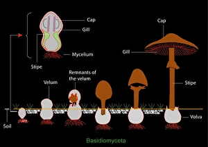
Mushroom anatomy. Diagram of the anatomy of Basidiomyceta mushrooms, and their growth from a fungal mycelium in the soil (left) to a full-grown mushroom (right)

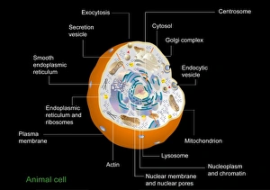
Background imageImages Dated 28th February 2011: Animal cell anatomy, diagram

Animal cell anatomy, diagram
Animal cell anatomy. Diagram showing the internal and external anatomy of an animal cell

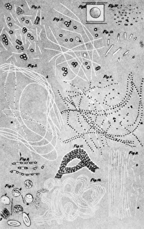
Background imageImages Dated 28th February 2011: Anthrax cultures, historical diagram

Anthrax cultures, historical diagram
Anthrax cultures. 1876 diagram of Bacillus anthracis (Anthrax) bacteria cultured by Robert Koch. Along with Louis Pasteur, Koch is considered the founder of modern medical bacteriology

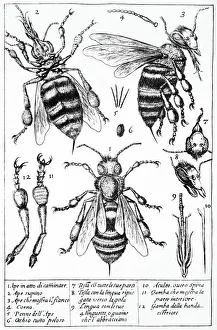
Background imageImages Dated 28th February 2011: Bee anatomy, historical artwork

Bee anatomy, historical artwork
Bees anatomy. 17th Century artwork by the Italian microscopist Francesco Stelluti, showing the anatomy of bees as seen by him under a microscope

Background imageImages Dated 28th February 2011: Louis Pasteur, French microbiologist

Louis Pasteur, French microbiologist
Louis Pasteur (1822-1895), French microbiologist and chemist, in his laboratory. Louis Pasteur proved the germ theory of disease, which holds that germs attack the body from the outside

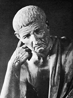
Background imageImages Dated 28th February 2011: Aristotle

Aristotle. Image of a sculpture in the Spada Gallery, Rome, of the Ancient Greek philosopher and naturalist Aristotle (384-322 BC)

Background imageImages Dated 28th February 2011: Antibiotic action, historical image

Antibiotic action, historical image
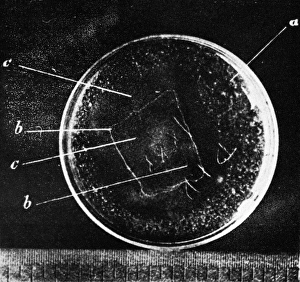
Antibiotic action. Historical image taken by the German pathologist Karl Dohle (1855-1928) in 1889, showing the action of an antibiotic compound on Bacillus anthracis (anthrax)

Background imageImages Dated 28th February 2011: Karl Rudolphi, Swedish naturalist

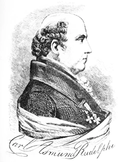
Karl Rudolphi, Swedish naturalist. Historical artwork of Karl Asmund Rudolphi (1771-1832). Rudolphi is credited with being the father of helminthology, the study of worms

Background imageImages Dated 28th February 2011: Tapeworm, historical artwork

Tapeworm, historical artwork. First recording of the Lumbricus latus tapeworm, drawn by the Flemish anatomist Adriaan van den Spiegel (aka Adrianus Spigelius) in 1618

Background imageImages Dated 28th February 2011: Disinfecting a house, historical artwork

Disinfecting a house, historical artwork
Disinfecting a house. 1734 artwork by Rene Antoine Ferchault de Reaumur depicting people in the 18th Century disinfecting a house to prevent infection

Background imageImages Dated 28th February 2011: Avicenna, Islamic physician

Avicenna, Islamic physician
Avicenna (980-1037). 17th Century artwork of the Islamic Persian physician and philosopher Avicenna (upper left) speaking to a group of students

Background imageImages Dated 28th February 2011: Aspirin, molecular structure diagram

Aspirin, molecular structure diagram
Aspirin. Diagram showing the molecular structure (left) and the chemical structure (right) of the painkilling drug aspirin (acetylsalycilic acid)

Background imageImages Dated 28th February 2011: 17th Century microscope, artwork

17th Century microscope, artwork
17th Century microscope. Historical artwork of a microscope built in the second half of the 17th Century by the Italian watchmaker and optician Giuseppe Campani (1635-1715)

Background imageImages Dated 28th February 2011: 18th Century scientist at work, artwork

18th Century scientist at work, artwork
18th Century scientist at work. 1782 artwork by Werner depicting an 18th Century scientist in his microscopy laboratory. Image from La Conquete Du Monde Invisible by Giuseppe Penso

Background imageImages Dated 28th February 2011: Edoardo Perroncito, Italian physician

Edoardo Perroncito, Italian physician
Edoardo Perroncito (1847Ôé¼" 1936), Italian physician, veterinarian and parasitologist. Perroncito is credited, along with Luigi Pagliani (1847Ôé¼" 1931)

Background imageImages Dated 28th February 2011: Historical microscope, artwork

Historical microscope, artwork
Historical microscope. 18th Century artwork of a microscope designed to observe aquatic organisms. This device was invented by the Swiss naturalist Abraham Trembley (1710Ôé¼" 1784) in 1747

Background imageImages Dated 28th February 2011: Bowmans capsule, diagram

Bowmans capsule, diagram
Bowmans capsule. Diagram showing the anatomical structure of a Bowmans capsule and glomerulus in a kidney

Background imageImages Dated 28th February 2011: Brain anatomy, artwork

Brain anatomy, artwork
Brain anatomy. Artwork of the underside (ventral aspect) of the brain, showing the anatomical structure of the various components

Background imageImages Dated 28th February 2011: Arthropod anatomy, diagram

Arthropod anatomy, diagram
Arthropod anatomy. Diagram showing the main anatomical parts of an arthropod, a large phylum of animals with this same basic body plan

Background imageImages Dated 28th February 2011: Vestibular ciliated cells, diagram

Vestibular ciliated cells, diagram
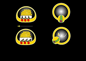
Vestibular ciliated cells. Diagram of the anatomical structure and function of the vestibular ciliated cells found in the inner ear that help sense and control of balance

Background imageImages Dated 28th February 2011: Protozoa, historical artwork

Protozoa, historical artwork
Protozoa. 1839 artwork by the naturalist Christian Gottfried Ehrenberg of Protozoa as seen under his microscope

Background imageImages Dated 28th February 2011: Hua Tuo, Chinese physician, artwork

Hua Tuo, Chinese physician, artwork
Hua Tuo, Chinese physician. Historical Chinese engraving depicting Hua Tuo (died circa 208 AD) standing by a tree. Hua Tuo was a famous Chinese physician who lived during the Han Dynasty

Background imageImages Dated 28th February 2011: Bacteriophage, diagram

Bacteriophage, diagram
Bacteriophage. Diagram of the anatomical structure of a bacteriophage, a virus that targets bacteria. The head (top) contains the DNA (red stands) within the capsid (blue hexagon)

Background imageImages Dated 28th February 2011: Human parasites, historical artwork

Human parasites, historical artwork
Human parasites. 1575 artwork of human parasites drawn by the Dutch physician, astronomer and astrologer Cornelius Gemma (1535Ôé¼" 1579)

Background imageImages Dated 28th February 2011: Brain anatomy, diagram

Brain anatomy, diagram
Brain anatomy. Diagram of the underside (ventral aspect) of the brain, showing the anatomical structure of the various components

Background imageImages Dated 28th February 2011: Meteorite found in France

Meteorite found in France
19th century hand coloured engraving of a meteorite, or aerolite, found at Juvenas in the Ardeche, the 5th June 1821. Reproduced from The Heavens by Amedee Guillemin, Richard Bentley 1878

Background imageImages Dated 28th February 2011: Milk-producing cell, artwork

Milk-producing cell, artwork
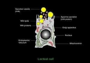
Milk-producing cell. Artwork of the anatomical structure of a lacteal (milk-producing) cell. Normal cell organelles include the nucleus (black, centre), the golgi apparatus (white)

Background imageImages Dated 28th February 2011: Bacteriorhodopsin, diagram

Bacteriorhodopsin, diagram
Bacteriorhodopsin. Diagram of the molecular structure of bacteriorhodopsin, a protein found in primitive micro-organisms known as Archaea

Background imageImages Dated 28th February 2011: Taste bud anatomy, diagram

Taste bud anatomy, diagram
Taste bud anatomy. Diagram of the anatomical structure of a taste bud on a tongue. The surface of the tongue is at bottom, with the tastebud (rounded structure) at centre

Background imageImages Dated 28th February 2011: Atheroma formation, diagram

Atheroma formation, diagram
Atheroma formation. Diagram showing the stages in the formation of an atheroma, a fatty plaque that forms on the inners walls of blood vessels in cardiovascular disease

Background imageImages Dated 28th February 2011: Ehrlichs side-chain theory, artwork

Ehrlichs side-chain theory, artwork. 1900 diagram by the German immunologist Paul Ehrlich (1854-1915) to explain his side-chain theory of immune response

Background imageImages Dated 28th February 2011: Semicircular canal, artwork

Semicircular canal, artwork
Semicircular canal. Artwork of the anatomical structure of the semicircular duct or canal, a structure in the inner ear that help detect head movements

Background imageImages Dated 28th February 2011: Semicircular canal, diagram

Semicircular canal, diagram
Semicircular canal. Diagram of the anatomical structure of the semicircular duct or canal, a structure in the inner ear that help detect head movements

Background imageImages Dated 28th February 2011: Paul Ehrlich, German immunologist

Paul Ehrlich, German immunologist
Paul Ehrlich (1854-1915), German immunologist. Ehrlich worked with aniline dyes, developing new methods of staining white blood cells. He discovered a new type of white blood cell, called mast cells

Background imageImages Dated 28th February 2011: Vestibular ciliated cells, artwork

Vestibular ciliated cells, artwork
Vestibular ciliated cells. Artwork of the anatomical structure and function of the vestibular ciliated cells found in the inner ear that help sense and control of balance

Background imageImages Dated 28th February 2011: Bacteriorhodopsin, artwork

Bacteriorhodopsin, artwork
Bacteriorhodopsin. Artwork of the molecular structure of bacteriorhodopsin, a protein found in primitive micro-organisms known as Archaea

Background imageImages Dated 28th February 2011: Protozoa, historical artwork

Protozoa, historical artwork
Protozoa. 1839 artwork by the naturalist Christian Gottfried Ehrenberg of Protozoa as seen under his microscope

Background imageImages Dated 28th February 2011: Elie Metchnikoff, Russian biologist

Elie Metchnikoff, Russian biologist
Elie Metchnikoff (1845-1916), Russian biologist and Nobel laureate. Metchnikoff, also known as Ilya Ilyich Mechnikov, is best known for his pioneering research into the immune system

Background imageImages Dated 28th February 2011: Gustavus Katterfelto, Prussian conjurer

Gustavus Katterfelto, Prussian conjurer
Gustavus Katterfelto (circa 1743-1799). Historical image of the Prussian conjurer, scientific lecturer and quack, Christian William Anthony Katterfelto

Background imageImages Dated 28th February 2011: Axon anatomy, diagram

Axon anatomy, diagram
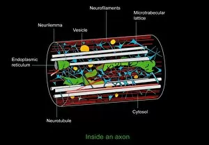
Axon anatomy. Diagram of the anatomical structure of an axon, the main extension (dendrite) of a nerve cell. Cellular components include endoplasmic reticulum (green), neurotubules (white)

Background imageImages Dated 28th February 2011: Capillary, artwork

Capillary, artwork
Capillary. Artwork showing the anatomical structure of a capillary seen in cross-section

Background imageImages Dated 28th February 2011: 18th Century entomology, artwork

18th Century entomology, artwork
18th Century entomology. Historical artwork by Rene Antoine Ferchault de Reaumur (1734) depicting 18th Century scientists catching insects for scientific study

Background imageImages Dated 28th February 2011: Retinal rod cell anatomy, artwork

Retinal rod cell anatomy, artwork
Retinal rod cell anatomy. Artwork of the external (left) and internal (right) anatomical structure of a retinal cell. These cells are found in the retina that lines the back of the eyes

Background imageImages Dated 28th February 2011: Preparing a culture, historical artwork

Preparing a culture, historical artwork
Preparing a culture. 1838 artwork by the naturalist Christian Gottfried Ehrenberg of a precursor to todays Petri dish, showing how to prepare a bacterial culture by swabbing the culture medium with a

Background imageImages Dated 28th February 2011: Girolamo Fracastoro, Italian physician

Girolamo Fracastoro, Italian physician
Girolamo Fracastoro (1478-1553), Italian physician and philosopher, historical artwork. Fracastoro proposed an early theory of the germ origin of disease

Background imageImages Dated 28th February 2011: 17th Century microscope, artwork

17th Century microscope, artwork
17th Century microscope. Historical artwork showing the construction of an early microscope. Image from La Conquete Du Monde Invisible by Giuseppe Penso

Background imageImages Dated 28th February 2011: Milk-producing cell, diagram

Milk-producing cell, diagram
Milk-producing cell. Diagram of the anatomical structure of a lacteal (milk-producing) cell. Normal cell organelles include the nucleus (black, centre), the golgi apparatus (white)

Background imageImages Dated 28th February 2011: Glomerular anatomy, diagram

Glomerular anatomy, diagram
Glomerular anatomy. Diagram of the anatomical structure of a glomerulus in a kidney


All Professionally Made to Order for Quick Shipping


-

Why Choose Us?

We are a leading provider of Art Prints and Photo Gifts since 2004, working in partnership with a range of Sporting Clubs, Charities, Museums and Picture Libraries. A large share of profits from any of their images will go directly towards supporting that charity or club. Our archive of images is carefully curated to bring you a wide range of subjects, including landscapes, wildlife, architecture, and more. We ship from our partner labs in the UK, USA, EU (Netherlands) and Australia.
+

How do I place an order?

Ordering is quick & easy - Just follow 5 Simple Steps:
  1. Find your image: Use our search box or browse our online photo Collections to find the image you want.

  2. Choose your print format: Select your desired product and add it to your cart.

  3. Enter your details: If you're a returning customer, simply enter your email address and password, and we'll fill in your billing and shipping address details. All personal details are held securely and are fully GDPR compliant. As standard, we remove all Personally Identifiable Information after 12 months.

  4. Pay for your purchase: We use state-of-the-art security for online shopping and do not have access to your card details.

  5. Sit back and relax: We'll email you confirmation of your order and when it's dispatched. Registered customers can also track orders in the 'My Account' area.

+

How do I pay for an order?

You can pay for your order with most of the major credit and debit cards, or PayPal. For added security, major financial institutions process payment details separately and securely on our behalf. We do not have access to your online payment card details. Online payments are preferred however we do take phone orders. For UK customers only we accept cheques issued against a UK bank.
+

Is my data safe?

We take data security very seriously. We do not have access to your full card details and all payments are requested over a fully secure connection. Additionally, we fully comply with current European and GDPR legislation, and automatically remove all personal data after at most 12 months (unless you have an account with us and have signed in recently). We also have a strict opt-in policy and would never sell your personal details. Your data is only used to fulfill your order promptly and efficiently.
+

Is wall art ready to hang?

For quick and easy installation all wall art, including framed prints, canvas prints and metal prints are supplied with a ready to hang solution on the back. Generally, saw tooth hangers are applied as they allow wall art to hang flush against the wall. The serrated edge of the hanger prevents the frame from shifting or tilting when hung.
+

Are the photo prints fade resistant?

Yes, we use archival quality photo paper photographic paper for vivid reproduction Prints are an accurate representations of the original artwork, which is preserved for artistic character and authenticity. We guarantee they match previews shown on our web site