Fraud Blocker Skip to main content

Home > Science Photo Library > Images Dated > 2009 > September > 16 Sep 2009

Images Dated 16th September 2009

Choose a picture from our Images Dated 16th September 2009 Collection for your Wall Art and Photo Gifts

49 items

We are proud to offer this selection in partnership with Science Photo Library

Background imageImages Dated 16th September 2009: Chaetognath, SEM

Chaetognath, SEM
Chaetognath. Scanning electron micrograph (SEM) of a chaetognath (phylum Chaetognatha). This microscopic predatory worm is transparent or translucent and is covered by a cuticle

Background imageImages Dated 16th September 2009: Maggot skin, SEM

Maggot skin, SEM
Maggot skin, coloured scanning electron micrograph (SEM). Maggots are the legless larvae of flies (order Diptera). Here, the skin of the maggot is covered in fine hooks which enables it to grip a

Background imageImages Dated 16th September 2009: Organic food, conceptual image

Organic food, conceptual image. Cut-out diagram of a mans head and digestive system, attached to a wooden background. Fruit and vegetables, and plastic sheep and pigs

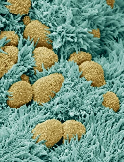
Background imageImages Dated 16th September 2009: Fallopian tube cells, SEM

Fallopian tube cells, SEM
Brain lining. Coloured scanning electron micrograph (SEM) of the lining of the brain, showing ependymal cells (yellow) and ciliary hairs (green)

Background imageImages Dated 16th September 2009: Womans face, conceptual artwork

Womans face, conceptual artwork. Abstract parts of a womans face (eyes, nose and mouth) on a background of polarised light effects. A red rose is at lower left

Background imageImages Dated 16th September 2009: Global environment, conceptual artwork

Global environment, conceptual artwork
Global environment, conceptual computer artwork. A spinning Earth globe is balanced on the extended finger of a plaster hand

Background imageImages Dated 16th September 2009: Mechanical technology, conceptual artwork

Mechanical technology, conceptual artwork
Mechanical technology, conceptual computer artwork. Rusty cogs and a timing dial are seen either side of a stepped graph showing an presentation of percentages

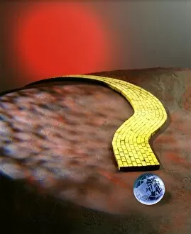
Background imageImages Dated 16th September 2009: Climate change, conceptual artwork

Climate change, conceptual artwork
Climate change, conceptual computer artwork. The Earth (lower right) is a small blue globe at the start of a yellow brick road in the shape of a partially visible question mark

Background imageImages Dated 16th September 2009: Climate change, conceptual artwork

Climate change, conceptual artwork
Climate change, conceptual computer artwork. The Earth (lower right) is a small globe on a background of sandy desert soil

Background imageImages Dated 16th September 2009: Ecological heart, conceptual image

Ecological heart, conceptual image. Germinating potatoes on a heart-shaped stone (centre). There is a network of copper pipes behind this, and leaves twining around the pipes

Background imageImages Dated 16th September 2009: Radioactive globe, conceptual artwork

Radioactive globe, conceptual artwork
Radioactive globe, conceptual computer artwork. The Earth (lower right) forms the centre of a radiation symbol. This image could represent events like a nuclear war

Background imageImages Dated 16th September 2009: Glass of water

Glass of water covered in water droplets, against a blue background

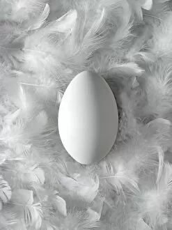
Background imageImages Dated 16th September 2009: Egg on feathers, conceptual image

Egg on feathers, conceptual image. This is a birds egg, surrounded by white bird feathers. The image represents concepts such as softness and fragility

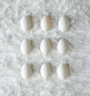
Background imageImages Dated 16th September 2009: Eggs on feathers, conceptual image

Eggs on feathers, conceptual image. These nine bird eggs, arrayed in a grid, are lying on a bed of white bird feathers. This image can represent the purity of mathematical concepts such as square

Background imageImages Dated 16th September 2009: Cosmic man juggling worlds, artwork

Cosmic man juggling worlds, artwork
Cosmic man juggling worlds. Conceptual computer artwork of a figure illuminated by polarised light, standing on the Earth and juggling four globes. The background is a starry night sky

Background imageImages Dated 16th September 2009: Wasp foot, SEM

Wasp foot, SEM
Wasp foot. Coloured scanning electron micrograph (SEM) of the foot of a common wasp (Vespula vulgaris). This is the final segment of the leg, known as the tarsus

Background imageImages Dated 16th September 2009: Maggot skin, SEM

Maggot skin, SEM
Maggot skin, coloured scanning electron micrograph (SEM). Maggots are the legless larvae of flies (order Diptera). Here, the skin of the maggot is covered in fine hooks which enables it to grip a

Background imageImages Dated 16th September 2009: Grape stem, light micrograph

Grape stem, light micrograph
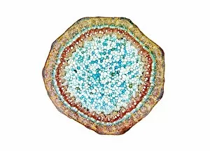
Grape stem. Light micrograph of a transverse section through a grape vine (Vitis sp.) stem. At the centre of the stem is a large area of pith, consisting of parenchyma cells

Background imageImages Dated 16th September 2009: Grape root, light micrograph

Grape root, light micrograph
Grape root. Light micrograph of a transverse section through a grape vine (Vitis sp.) root. At centre are xylem (red) and tracheid (green) cells, which transport water around the plant

Background imageImages Dated 16th September 2009: Compound eye of a gnat, SEM

Compound eye of a gnat, SEM
Compound eye of a gnat. Scanning electron micrograph (SEM) of the surface of a compound eye from a fungus gnat (suborder Nematocera). The eye consists of many rounded lenses known as ommatidia

Background imageImages Dated 16th September 2009: Sweet potato stem, light micrograph

Sweet potato stem, light micrograph
Sweet potato stem. Light micrograph of a transverse section through part of a sweet potato (Ipomoea batatas) stem. At bottom is a large area of pith, consisting of parenchyma cells

Background imageImages Dated 16th September 2009: Sweet potato stem, light micrograph

Sweet potato stem, light micrograph
Sweet potato stem. Light micrograph of a transverse section through a sweet potato (Ipomoea batatas) stem. At the centre of the stem is a large area of pith, consisting of parenchyma cells

Background imageImages Dated 16th September 2009: Head of a pipistrelle bat

Head of a pipistrelle bat. Coloured scanning electron micrograph (SEM) of the head of a pipistrelle bat (Pipistrellus pipistrellus)

Background imageImages Dated 16th September 2009: Sweet potato root, light micrograph

Sweet potato root, light micrograph
Sweet potato root. Light micrograph of a transverse section through the centre of a sweet potato (Ipomoea batatas) root. Xylem cells, which transport water around the plant, are red

Background imageImages Dated 16th September 2009: Sweet potato root, light micrograph

Sweet potato root, light micrograph
Sweet potato root. Light micrograph of a transverse section through a sweet potato (Ipomoea batatas) root. At centre are xylem cells (red), which transport water around the plant

Background imageImages Dated 16th September 2009: Water vapour distribution, computer model

Water vapour distribution, computer model. Coloured supercomputer simulations showing the distribution of the Earths water vapour. Atmospheric water vapour is invisible

Background imageImages Dated 16th September 2009: Clematis stem, light micrograph

Clematis stem, light micrograph
Clematis stem. Light micrograph of a transverse section through the stem of a clematis (Clematis flammula) plant. At the centre of the stem is a large area of pith, consisting of parenchyma cells

Background imageImages Dated 16th September 2009: Sweet potato leaf, light micrograph

Sweet potato leaf, light micrograph
Sweet potato leaf. Light micrograph of a transverse section through the midrib of a sweet potato (Ipomoea batatas) leaf. The semicircular structure at centre is the vascular bundle

Background imageImages Dated 16th September 2009: Clematis stem, light micrograph

Clematis stem, light micrograph
Clematis stem. Light micrograph of a transverse section through the stem of a clematis (Clematis flammula) plant. At the centre of the stem is a large area of pith, consisting of parenchyma cells

Background imageImages Dated 16th September 2009: White bryony stem, light micrograph

White bryony stem, light micrograph
White bryony stem. Light micrograph of a transverse section through the stem of a white bryony (Bryonia dioica) stem. At the centre of the stem is the pith, consisting of parenchyma cells

Background imageImages Dated 16th September 2009: Diatoms, SEM

Diatoms, SEM
Diatoms. Coloured scanning electron micrograph (SEM) of diatoms (blue) attached to algae (brown). The diatoms are a group of photosynthetic, single-celled algae containing about 100, 000 species

Background imageImages Dated 16th September 2009: Diatoms, SEM

Diatoms, SEM
Diatoms. Coloured scanning electron micrograph (SEM) of diatoms (red) attached to algae (grey). The diatoms are a group of photosynthetic, single-celled algae containing about 100, 000 species

Background imageImages Dated 16th September 2009: Diatom, SEM

Diatom, SEM
Diatom. Coloured scanning electron micrograph (SEM) of the wall of the diatom Thalassiosira sp.. Thalassiosira is an ocean diatom that is a major participant in the biological pumping of carbon to

Background imageImages Dated 16th September 2009: Diatom, SEM

Diatom, SEM
Diatom. Coloured scanning electron micrograph (SEM) of the wall of the diatom Thalassiosira sp.. Thalassiosira is an ocean diatom that is a major participant in the biological pumping of carbon to

Background imageImages Dated 16th September 2009: Starch grains, light micrograph

Starch grains, light micrograph
Starch grains. Polarised light micrograph of starch grains from a lily (Canna sp.) root tuber. Starch is used as a source of energy for a growing plant

Background imageImages Dated 16th September 2009: Liana stem, light micrograph

Liana stem, light micrograph
Liana stem. Polarised light micrograph of a transverse section through the stem of a Brazilian liana, or woody vine. The tissues within this stem have differentiated to form many secondary stem-like

Background imageImages Dated 16th September 2009: Liana stem, light micrograph

Liana stem, light micrograph
Liana stem. Polarised light micrograph of a transverse section through the stem of a Brazilian liana, or woody vine. At the centre of the stem is the pith, consisting of parenchyma cells

Background imageImages Dated 16th September 2009: Liana stem, light micrograph

Liana stem, light micrograph
Liana stem. Light micrograph of a transverse section through the stem of a liana (Aristolochia tormentosa), or woody vine. At the centre of the stem is the pith, consisting of parenchyma cells

Background imageImages Dated 16th September 2009: Bat tick mouthparts, SEM

Bat tick mouthparts, SEM
Bat tick mouthparts. Coloured scanning electron micrograph (SEM) of a bat tick (order Acarina), an arachnid ectoparasite (external parasites) that feeds on the blood of bats

Background imageImages Dated 16th September 2009: Liana stem, light micrograph

Liana stem, light micrograph
Liana stem. Light micrograph of a transverse section through the stem of a liana (Aristolochia sp.), or woody vine. At the centre of the stem is the pith, consisting of parenchyma cells

Background imageImages Dated 16th September 2009: Bat tick mouthparts, SEM

Bat tick mouthparts, SEM
Bat tick mouthparts. Coloured scanning electron micrograph (SEM) of a bat tick (order Acarina), an arachnid ectoparasite (external parasites) that feeds on the blood of bats

Background imageImages Dated 16th September 2009: Rosemary leaf, light micrograph

Rosemary leaf, light micrograph
Rosemary leaf. Light micrograph of a transverse section through a rosemary (Romarinus officinalis) leaf. The leaf of this plant has a number of adaptations that help it to minimise water loss through

Background imageImages Dated 16th September 2009: Woven silk, SEM

Woven silk, SEM
Woven silk, coloured scanning electron micrograph (SEM). Silk is a protein fibre, which can be woven into textiles. It is obtained from the cocoons of metamorphosising silkworms

Background imageImages Dated 16th September 2009: Woven silk, SEM

Woven silk, SEM
Woven silk, coloured scanning electron micrograph (SEM). Silk is a protein fibre, which can be woven into textiles. It is obtained from the cocoons of metamorphosising silkworms

Background imageImages Dated 16th September 2009: Pincushion leaf, light micrograph

Pincushion leaf, light micrograph
Pin cushion leaf. Polarised light micrograph of a transverse section through a pinchusion (Hakea laurina) leaf. The leaf of this plant has a number of adaptations that help it to minimise water loss

Background imageImages Dated 16th September 2009: Woven silk, SEM

Woven silk, SEM
Woven silk, coloured scanning electron micrograph (SEM). Silk is a protein fibre, which can be woven into textiles. It is obtained from the cocoons of metamorphosising silkworms


All Professionally Made to Order for Quick Shipping


-

Why Choose Us?

We are a leading provider of Art Prints and Photo Gifts since 2004, working in partnership with a range of Sporting Clubs, Charities, Museums and Picture Libraries. A large share of profits from any of their images will go directly towards supporting that charity or club. Our archive of images is carefully curated to bring you a wide range of subjects, including landscapes, wildlife, architecture, and more. We ship from our partner labs in the UK, USA, EU (Netherlands) and Australia.
+

How do I place an order?

Ordering is quick & easy - Just follow 5 Simple Steps:
  1. Find your image: Use our search box or browse our online photo Collections to find the image you want.

  2. Choose your print format: Select your desired product and add it to your cart.

  3. Enter your details: If you're a returning customer, simply enter your email address and password, and we'll fill in your billing and shipping address details. All personal details are held securely and are fully GDPR compliant. As standard, we remove all Personally Identifiable Information after 12 months.

  4. Pay for your purchase: We use state-of-the-art security for online shopping and do not have access to your card details.

  5. Sit back and relax: We'll email you confirmation of your order and when it's dispatched. Registered customers can also track orders in the 'My Account' area.

+

How do I pay for an order?

You can pay for your order with most of the major credit and debit cards, or PayPal. For added security, major financial institutions process payment details separately and securely on our behalf. We do not have access to your online payment card details. Online payments are preferred however we do take phone orders. For UK customers only we accept cheques issued against a UK bank.
+

Is my data safe?

We take data security very seriously. We do not have access to your full card details and all payments are requested over a fully secure connection. Additionally, we fully comply with current European and GDPR legislation, and automatically remove all personal data after at most 12 months (unless you have an account with us and have signed in recently). We also have a strict opt-in policy and would never sell your personal details. Your data is only used to fulfill your order promptly and efficiently.
+

Is wall art ready to hang?

For quick and easy installation all wall art, including framed prints, canvas prints and metal prints are supplied with a ready to hang solution on the back. Generally, saw tooth hangers are applied as they allow wall art to hang flush against the wall. The serrated edge of the hanger prevents the frame from shifting or tilting when hung.
+

Are the photo prints fade resistant?

Yes, we use archival quality photo paper photographic paper for vivid reproduction Prints are an accurate representations of the original artwork, which is preserved for artistic character and authenticity. We guarantee they match previews shown on our web site