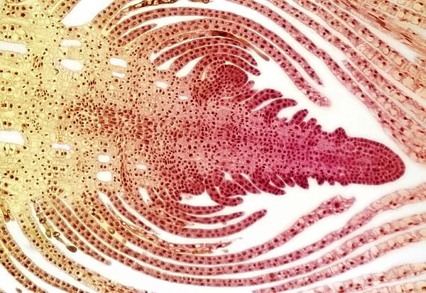
Hydrilla bud, light micrograph
![]()

Wall Art and Photo Gifts from Science Photo Library
Hydrilla bud, light micrograph
Hydrilla bud. Light micrograph of a longitudinal section through an axillary bud on a Hydrilla plant, showing its internal structure. Hydrilla is a genus of aquatic plant native to the warmer parts of Asia and adapted to life in submerged freshwater environments. Seen here is a bud, an undeveloped embryonic shoot, with the apical meristem, or growing tip, at centre right (red). This is undifferentiated meristemic tissue, which lays down a growing root or shoot behind itself, pushing itself forward. Behind this (to the left) are leaf primordia (young leaves), which have recently formed
Science Photo Library features Science and Medical images including photos and illustrations
Media ID 6291055
© STEVE GSCHMEISSNER/SCIENCE PHOTO LIBRARY
Apical Meristem Axillary Fresh Water Growing Growth Histological Histology Internal Longitudinal Root Scale Scales Shoot Tissue Undifferentiated Young Cells Light Micrograph Light Microscope Section Sectioned
EDITORS COMMENTS
This print showcases the intricate beauty of a Hydrilla bud, captured through a light micrograph. The image reveals the internal structure of an axillary bud on a Hydrilla plant, providing insight into its fascinating nature. Hydrilla is an aquatic plant genus that thrives in submerged freshwater environments, primarily found in warmer regions of Asia. In this snapshot, we witness the essence of growth and development as an undeveloped embryonic shoot takes center stage with its apical meristem prominently displayed in red hues. This undifferentiated meristemic tissue serves as the driving force behind root or shoot formation by continuously pushing itself forward. Adjacent to the apical meristem lie leaf primordia - young leaves that have recently formed. These delicate structures add depth and texture to the composition, showcasing their potential for future growth and contribution to the overall vitality of the plant. The image provides a glimpse into the microscopic world within plants, highlighting their cellular complexity and biological processes. It offers valuable insights into botany, biology, and histology while celebrating nature's remarkable ability to adapt and thrive even in challenging environments. Captured using a light microscope at high magnification levels, this sectioned view allows us to appreciate every detail with precision. Science Photo Library has once again provided us with an awe-inspiring visual representation that reminds us of both our curiosity about life's mysteries and our appreciation for its inherent beauty.
MADE IN THE USA
Safe Shipping with 30 Day Money Back Guarantee
FREE PERSONALISATION*
We are proud to offer a range of customisation features including Personalised Captions, Color Filters and Picture Zoom Tools
SECURE PAYMENTS
We happily accept a wide range of payment options so you can pay for the things you need in the way that is most convenient for you
* Options may vary by product and licensing agreement. Zoomed Pictures can be adjusted in the Cart.

