Home > Science > SEM
Honey bee mite, SEM

![]()
Wall Art and Photo Gifts from Science Photo Library
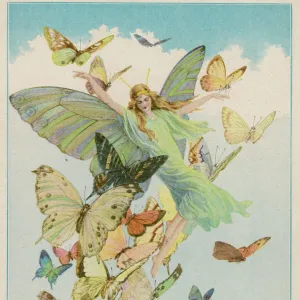
Honey bee mite, SEM
Honey bee mite. Coloured scanning electron micrograph (SEM) of a honey bee mite (Varroa sp.). Parasitic mites have decimated hives of wild and domesticated honey bees (Apis mellifera) throughout the world. Low levels of infestation are difficult to detect, but are often fatal to the colony within three to seven years. Mite eggs are laid in a hives brood cells. Mite larvae feed on the developing bee larvae, mate and then leave the cell. Female mites attach themselves to the body of an adult worker or drone bee and feed from it. When ready to lay her eggs she will drop into the hives brood cells and complete the cycle. Magnification: x60 when printed 10 centimetres wide
Science Photo Library features Science and Medical images including photos and illustrations
Media ID 6465781
© STEVE GSCHMEISSNER/SCIENCE PHOTO LIBRARY
Body Deadly Ecto Parasite External False Colour Fatal Honey Bee Legs Mite Parasite Parasitic Parasitising Surface False Coloured Honey Bee Mite
EDITORS COMMENTS
This print showcases the intricate world of a honey bee mite, captured through a coloured scanning electron micrograph (SEM). These parasitic mites have wreaked havoc on both wild and domesticated honey bees worldwide. Although low levels of infestation may go unnoticed, they can prove fatal to entire colonies within three to seven years. The life cycle of these tiny pests begins with the laying of eggs in the brood cells of beehives. The mite larvae then feed on developing bee larvae before maturing and leaving their host cell. Female mites attach themselves to adult worker or drone bees, feeding off them until they are ready to lay their own eggs. Once prepared, they drop into the brood cells again, completing this vicious cycle. At a magnification of x60 when printed at 10 centimetres wide, this image reveals the astonishing details present on the surface of these parasites' bodies and legs. The false colouring adds an artistic touch while highlighting essential biological features under scrutiny by researchers using scanning electron microscopes. This photograph not only offers insight into the fascinating world of honey bee mites but also serves as a reminder of their deadly impact on vital pollinators like Apis mellifera. It stands as a testament to our ongoing efforts in understanding and combating these ectoparasites that threaten one of nature's most crucial species – honey bees.
MADE IN THE USA
Safe Shipping with 30 Day Money Back Guarantee
FREE PERSONALISATION*
We are proud to offer a range of customisation features including Personalised Captions, Color Filters and Picture Zoom Tools
SECURE PAYMENTS
We happily accept a wide range of payment options so you can pay for the things you need in the way that is most convenient for you
* Options may vary by product and licensing agreement. Zoomed Pictures can be adjusted in the Cart.