Home > Arts > Artists > P > those present
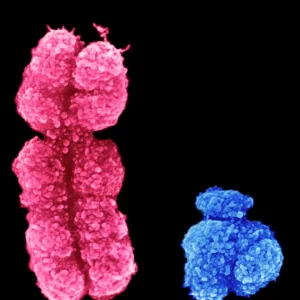
Coloured SEM of an embryo at the stage of morula

![]()
Wall Art and Photo Gifts from Science Photo Library
Coloured SEM of an embryo at the stage of morula
Embryo development. Coloured scanning electron micrograph of an embryo at the early stage known as the morula. The egg reaches this phase about 4 days after fertilisation after a series of mitotic divisions. At this stage about 12-16 cells are present and are surrounded by a thin glycoprotein layer, the zona pellucida, which was here removed. The inner cells of the morula will give rise to the tissues of the embryo while the outer cells, covered here by microvilli (tiny yellow ridges), will form the placenta. The morula will implant into the uterus six days after fertilisation. Magnification: x1350 at 6x7cm size
Science Photo Library features Science and Medical images including photos and illustrations
Media ID 6454257
© PROFESSORS P.M. MOTTA & J. VAN BLERKOM/ SCIENCE PHOTO LIBRARY
FEATURES IN THESE COLLECTIONS
> Arts
> Artists
> P
> those present
EDITORS COMMENTS
This print showcases the intricate beauty of an embryo at the early stage known as morula. Through the lens of a coloured scanning electron microscope, we are granted a glimpse into the fascinating world of embryonic development. Approximately four days after fertilisation and following a series of mitotic divisions, this morula emerges. Comprising 12-16 cells, it is encased within a delicate glycoprotein layer called the zona pellucida, which has been meticulously removed in this image. The inner cells hold within them the potential to give rise to various tissues that will form the foundation of life itself. Intriguingly, surrounding these vital inner cells are outer cells adorned with microvilli - tiny yellow ridges visible here. These outer cells play a crucial role in forming the placenta, ensuring nourishment and support for future growth and development. Six days after fertilisation, this remarkable morula will implant itself into its nurturing home - the uterus. This awe-inspiring journey marks just one small step in an extraordinary process that ultimately leads to new life. With magnification set at x1350 on a 6x7cm scale, Science Photo Library has captured not only scientific precision but also artistic brilliance in presenting us with this profound snapshot of human existence.
MADE IN THE USA
Safe Shipping with 30 Day Money Back Guarantee
FREE PERSONALISATION*
We are proud to offer a range of customisation features including Personalised Captions, Color Filters and Picture Zoom Tools
SECURE PAYMENTS
We happily accept a wide range of payment options so you can pay for the things you need in the way that is most convenient for you
* Options may vary by product and licensing agreement. Zoomed Pictures can be adjusted in the Cart.