Home > Science > SEM
Breathing tube on a fruit flys pupa, SEM
![]()

Wall Art and Photo Gifts from Science Photo Library
Breathing tube on a fruit flys pupa, SEM
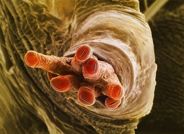
Breathing tube on a fruit flys pupa, coloured scanning electron micrograph (SEM). This structure is called a spiracle. This fruit fly is Drosophila melanogaster (wild type Oregon R), and its larva develop within a structure called a puparium, from which pupal spiracles project outward. The anterior pair of spiracles terminate in tubelike extensions, called spiracular papillae (red). The exposed ends open in narrow slits, which take in air and pass it to the developing pupa. Magnification: x470 when printed 10 centimetres wide
Science Photo Library features Science and Medical images including photos and illustrations
Media ID 6461722
© DR JEREMY BURGESS/SCIENCE PHOTO LIBRARY
Animal Body Anterior Breathing Tube Developing Developmental Biology Drosophila Melanogaster Entomological False Colour Front Fruit Fly Insecta Larval Stage Metamorphosis Oxygen Papilla Papillae Pupa Respiration Respiratory Spiracle Spiracles Tube Tubes False Coloured
EDITORS COMMENTS
This print showcases the intricate breathing tube of a fruit fly's pupa, captured using a coloured scanning electron microscope (SEM). Known as a spiracle, this structure plays a vital role in respiration. The featured fruit fly belongs to the Drosophila melanogaster species, specifically the wild type Oregon R variant. During its larval stage, this tiny insect develops within a protective casing called a puparium, from which these fascinating pupal spiracles emerge. The anterior pair of spiracles on display terminate in striking tubelike extensions known as spiracular papillae, highlighted in vibrant red hues. These papillae possess narrow slits at their exposed ends that allow air to enter and be passed on to the developing pupa. With an impressive magnification of x470 when printed 10 centimetres wide, this image offers us an up-close look at the complex anatomy and biological processes taking place within these remarkable creatures. As we delve into the world of entomology and explore nature's incredible diversity, images like these remind us of the beauty found even in minuscule organisms. They also serve as valuable tools for studying developmental biology and understanding metamorphosis among insects. This photograph is yet another testament to Science Photo Library's commitment to capturing awe-inspiring moments from our natural world without commercial intent but with utmost scientific precision.
MADE IN THE USA
Safe Shipping with 30 Day Money Back Guarantee
FREE PERSONALISATION*
We are proud to offer a range of customisation features including Personalised Captions, Color Filters and Picture Zoom Tools
SECURE PAYMENTS
We happily accept a wide range of payment options so you can pay for the things you need in the way that is most convenient for you
* Options may vary by product and licensing agreement. Zoomed Pictures can be adjusted in the Cart.

