Home > Architecture > Related Images
Difflugia Corona

![]()
Wall Art and Photo Gifts from Mary Evans Picture Library
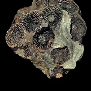
Difflugia Corona
Freshwater Testate Amoebae. Magnification x 450
Mary Evans Picture Library makes available wonderful images created for people to enjoy over the centuries
Media ID 8619615
© Mary Evans Picture Library 2015 - https://copyrighthub.org/s0/hub1/creation/maryevans/MaryEvansPictureID/10711988
Amoebozoa Case Electron Micrograph Eukaryote Eukaryotic Freshwater Grain Magnification Micrograph Microscope Microscope Image Protist Protista Protozoa Protozoan Sand Scanning Electron Micrograph Scanning Electron Microscope Scanning Electron Microscope Image Sediment Sem Image Amoebae
EDITORS COMMENTS
1. Title: Magnificent Architecture of Difflugia Corona: A Microscopic Exploration of Freshwater Testate Amoebae 2.. This stunning scanning electron micrograph (SEM) presents a captivating view of Difflugia Corona, a member of the Amoebozoa phylum, specifically the Arcellinida class, and the Coronate subclass. This eukaryotic protist, belonging to the Difflugiidae family, is commonly found in freshwater sediments. 3. Background: Difflugia Corona is renowned for its intricate, architectural test, which resembles a miniature house. The test, made primarily of calcium carbonate, provides the amoeba with protection and stability, enabling it to thrive in various aquatic environments. 4. Detailed Inspection: In this SEM image, the intricate details of the Difflugia Corona's test are revealed. The test exhibits a coronal structure, featuring a central, cylindrical body with a series of radiating arms. The surface of the test is adorned with tiny, granular structures, reminiscent of sand grains, which add to its architectural complexity. 5. Technique: The image was captured using a Scanning Electron Microscope (SEM) at a magnification of 450x. This advanced microscopy technique allows for the high-resolution visualization of the microscopic world, revealing intricate details that are otherwise invisible to the naked eye. 6. Significance: The discovery and study of Difflugia Corona and other testate amoebae contribute significantly to the fields of microbiology, ecology, and paleontology. These organisms provide valuable insights into the evolution of eukaryotic cells and the complex interplay between organisms and their environments. 7. Conclusion: This mesmerizing SEM image of Difflugia Corona not only showcases the beauty of the natural world but also highlights the importance of microscopy in expanding our understanding of the intricacies of life.
MADE IN THE USA
Safe Shipping with 30 Day Money Back Guarantee
FREE PERSONALISATION*
We are proud to offer a range of customisation features including Personalised Captions, Color Filters and Picture Zoom Tools
FREE COLORIZATION SERVICE
You can choose advanced AI Colorization for this picture at no extra charge!
SECURE PAYMENTS
We happily accept a wide range of payment options so you can pay for the things you need in the way that is most convenient for you
* Options may vary by product and licensing agreement. Zoomed Pictures can be adjusted in the Cart.