Home > Europe > United Kingdom > Wales > Flintshire > Mold
Aspergillus

![]()
Wall Art and Photo Gifts from Mary Evans Picture Library
Aspergillus
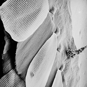
An SEM image of aspergillus in spore production (x 815 on a standard 9 cm wide print). The moulds are common in the northern hemisphere and some cause disease in humans and animals
Mary Evans Picture Library makes available wonderful images created for people to enjoy over the centuries
Media ID 8614097
© Mary Evans Picture Library 2015 - https://copyrighthub.org/s0/hub1/creation/maryevans/MaryEvansPictureID/10707003
Ascomycete Ascomycota Chain Disease Electron Electron Micrograph Fungi Fungus Linked Magnified Micrograph Microscope Image Mold Mould Scanning Scanning Electron Micrograph Scanning Electron Microscope Scanning Electron Microscope Image Sem Image Spore
EDITORS COMMENTS
1. Title: Magnified Majesty: An Aspergillus Ascomycete in Spore Production (SEM Image) 2. Description: Witness the intricate beauty and complexity of the natural world in this Scanning Electron Micrograph (SEM) image of Aspergillus, a common mold belonging to the Ascomycota phylum, specifically the Eurotiales order and the Trichocomaceae family. This micrograph, captured at a staggering magnification of 815x, reveals the intricacies of this ascomycete's spore-producing structures. 3. Background: Aspergillus is a ubiquitous genus of fungi found in various environments, particularly in the northern hemisphere. Some species of Aspergillus are beneficial, contributing to the decomposition of organic matter and the production of food and industrial enzymes. However, others are notorious for causing diseases in humans and animals, including aspergillosis and foodborne illnesses. 4. Details: In this image, the fungal structures appear as a chain-like arrangement, with each unit consisting of a cluster of asci (spore-bearing sacs) and their associated paraphyses (supporting structures). The asci are encased within a protective cover called the ascocarp, which is not visible at this magnification. The spores, which appear as small, round structures, are the reproductive units of the fungus, responsible for its spread and survival. 5. Technique: The image was captured using a Scanning Electron Microscope, a powerful tool that allows for the examination of surfaces with high resolution and depth of field. The sample was likely coated with a thin layer of conductive material to facilitate the flow of electrons and prevent charging during imaging. The resulting micrograph provides a fascinating glimpse into the microscopic world of Aspergillus and its spore production process.
MADE IN THE USA
Safe Shipping with 30 Day Money Back Guarantee
FREE PERSONALISATION*
We are proud to offer a range of customisation features including Personalised Captions, Color Filters and Picture Zoom Tools
FREE COLORIZATION SERVICE
You can choose advanced AI Colorization for this picture at no extra charge!
SECURE PAYMENTS
We happily accept a wide range of payment options so you can pay for the things you need in the way that is most convenient for you
* Options may vary by product and licensing agreement. Zoomed Pictures can be adjusted in the Cart.