Fraud Blocker Skip to main content

X Ray Collection (#21)

"Unveiling the Hidden World: Exploring the Marvels of X-ray Imaging" Pillars of Creation: Witness the breathtaking beauty of celestial formations through an X-ray lens

Background imageX Ray Collection: Normal female pelvis, X-ray

Normal female pelvis, X-ray
Normal female pelvis, coloured frontal X-ray. The thigh (femur) bones of the leg are seen at bottom left and right, and the base of the spine is at top

Background imageX Ray Collection: Lung abscess, X-ray

Lung abscess, X-ray
Lung abscess. Coloured X-ray showing an abscess (dense coloured area, upper right) in the upper lobe of a patients left lung (right). An abscess is an accumulation of puss within a cavity in tissue

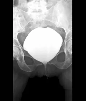
Background imageX Ray Collection: Full bladder, X-ray cystography

Full bladder, X-ray cystography
Full bladder. X-ray cystogram of the pelvic region of a 50-year-old incontinent patient, showing urine (white) leaking (lower centre) from the bladder (round, centre) during a stress test

Background imageX Ray Collection: Portable high resolution CT scanner

Portable high resolution CT scanner with fossil skull.This CT (computed tomography) scanner fires beams of X-rays (left) at different angles around the skull (centre)

Background imageX Ray Collection: Galaxy cluster collision, X-ray image

Galaxy cluster collision, X-ray image
Galaxy cluster MACS J0025.4-1222 collision. Combined Hubble Space Telescope (HST) and Chandra X-ray Observatory (CXO) image of a powerful collision of galaxy clusters

Background imageX Ray Collection: The First Attempt to Treat Cancer with X Rays by Doctor Chicotot, 1907 (oil on canvas)

The First Attempt to Treat Cancer with X Rays by Doctor Chicotot, 1907 (oil on canvas)
XIR23962 The First Attempt to Treat Cancer with X Rays by Doctor Chicotot, 1907 (oil on canvas) by Chicotot, Georges (fl.1889-1907); 130x85 cm; Musee de l Assistance Publique, Hopitaux de Paris

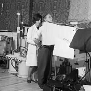
Background imageX Ray Collection: Medical screening in Rotherham, South Yorkshire, 1967. Artist: Michael Walters

Medical screening in Rotherham, South Yorkshire, 1967. Artist: Michael Walters
Medical screening in Rotherham, South Yorkshire, 1967. In a photograph taken for the Central Office of Information, a nurse supervises the taking of an X-ray of a patients chest as part of a health

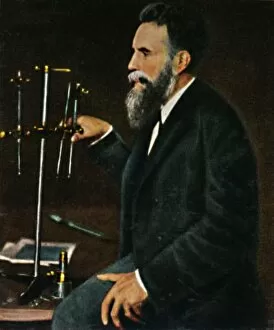
Background imageX Ray Collection: Wilhelm von Rontgen 1845-1923, 1934

Wilhelm von Rontgen 1845-1923, 1934. Wilhelm Conrad Rontgen (1845-1923), German mechanical engineer and physicist, who produced

Background imageX Ray Collection: Crookes, Rontgen and Finsen - Using the Marvellous X-Rays Apparatus, c1925

Crookes, Rontgen and Finsen - Using the Marvellous X-Rays Apparatus, c1925. Niels T. Finsen, (1860-1904). From Cassells Romance of Famous Lives, Volume 2 by Harold Wheeler

Background imageX Ray Collection: Interior of a Portland field hospital during the Boer War in South Africa, 1900

Interior of a Portland field hospital during the Boer War in South Africa, 1900. Artist: Anthony Bowlby
Interior of a Portland field hospital during the Boer War in South Africa. The X-rays and operating bed, 1900. From Black & White Budget, Vol. III. [The Black and White Publishing Company, J P

Background imageX Ray Collection: Major Battersby and his Orderly taking a Radiograph in the Soudan, c1890, (1910)

Major Battersby and his Orderly taking a Radiograph in the Soudan, c1890, (1910). From The Strand Magazine. [George Newnes Ltd. London, 1910]

Background imageX Ray Collection: X-ray apparatus used for examination of suspected cases of heart or lung disease, c1935 (c1937)

X-ray apparatus used for examination of suspected cases of heart or lung disease, c1935 (c1937)
X-ray apparatus used at the Central Medical Board for examination of suspected cases of heart or lung disease in prospective commercial pilots, c1935 (c1937). From Wonders of World Aviation, Vol

Background imageX Ray Collection: Million volt X-ray tube, 1938

Million volt X-ray tube, 1938. Churchmans cigarette card, from a series titled Modern Wonders [WA & AC Churchman, Great Britain & Ireland, 1938]

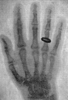
Background imageX Ray Collection: X-ray photograph of a hand, 1926

X-ray photograph of a hand, 1926. Also featured is an inset of Wilhelm Rontgen (1845-1913) who discovered x-rays in 1895. From An Outline of Christianity, The Story of Our Civilisation

Background imageX Ray Collection: The examination of a childs arm by radiography, 19th century. Artist: Gustave Francois Lasellaz

The examination of a childs arm by radiography, 19th century. Artist: Gustave Francois Lasellaz
The examination of a childs arm by radiography, 19th century. A print from les Dernieres Merveilles de la Science, by Daniel Bellet

Background imageX Ray Collection: Wilhelm Konrad von Rontgen, German physicist, 1902

Wilhelm Konrad von Rontgen, German physicist, 1902. In 1895, while professor of physics at Wurzburg, (Rontgen (1845-1913) discovered X-rays, originally called Rontgen rays

Background imageX Ray Collection: Examining a patients thorax using an X-ray tube and fluorescent screen, 1903

Examining a patients thorax using an X-ray tube and fluorescent screen, 1903. The X-ray tube (on tripod) is set at the required height and the patient stands directly in front of it

Background imageX Ray Collection: X-ray apparatus, 1915

X-ray apparatus, 1915. Apparatus powered by a Ruhmkorff coil being used to take an X-ray of a hand. Cigarette card

Background imageX Ray Collection: X-raying the hand, 1924

X-raying the hand, 1924. An X-ray tube and an X-ray photograph of a hand, with the bones and a wristwatch and ring clearly visible. Cigarette card

Background imageX Ray Collection: X-ray image of a solar flare

X-ray image of a solar flare. Solar flares, or prominences, are massive eruptions of ionized hydrogen gas, often larger than the Earth, which arch away from the surface of the Sun

Background imageX Ray Collection: X-ray image of sun, Skylab, 1970s

X-ray image of sun, Skylab, 1970s. This image shows a coronal hole. Skylab was launched on 14 May 1973 and was Americas first manned orbiting space station

Background imageX Ray Collection: Wilhelm Konrad von Roentgen, German physicist, 1901

Wilhelm Konrad von Roentgen, German physicist, 1901. The discover of X-rays, Roentgen was awarded the Nobel prize for Physics in 1901. Photograph courtesy of the Nobel Foundation

Background imageX Ray Collection: Discharge in Geissler tubes containing rarefied gases, 1887

Discharge in Geissler tubes containing rarefied gases, 1887
Discharge in Geissler tubes containing rarefied gases. Investigations of these phenomena led to the discovery of cathode rays, x-rays and electrons. From A. Ganot Natural Philosophy. (London, 1887)

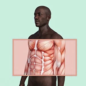
Background imageX Ray Collection: bald, body, chest, close up, color image, concept, condition, copy space, cut out

bald, body, chest, close up, color image, concept, condition, copy space, cut out
Chest and abdominal muscles in man

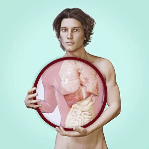
Background imageX Ray Collection: body, brunette, circular, close up, colon, color image, concept, copy space, cut out

body, brunette, circular, close up, colon, color image, concept, copy space, cut out
Internal organs of man

Background imageX Ray Collection: Illustration of doctor pointing at x-ray of pelvis and spine

Illustration of doctor pointing at x-ray of pelvis and spine

Background imageX Ray Collection: CHICAGO: SANITARIUM, 1941. Pneumothorax treatment for a patient with tuberculosis

CHICAGO: SANITARIUM, 1941. Pneumothorax treatment for a patient with tuberculosis at the municipal sanitarium on the South Side of Chicago, Illinois. Photograph by Russell Lee, 1941

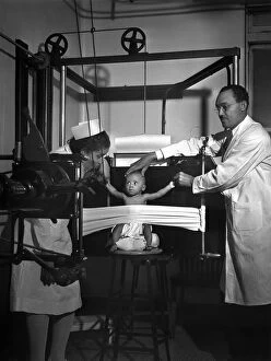
Background imageX Ray Collection: X-RAY MACHINE, 1942. A baby being x-rayed at the Provident Hospital in Chicago, Illinois

X-RAY MACHINE, 1942. A baby being x-rayed at the Provident Hospital in Chicago, Illinois. Photograph by Jack Delano in March 1942

Background imageX Ray Collection: X-RAY ROOM, c1920. An X-ray room at the Washington Sanitarium in Takoma Park, Maryland

X-RAY ROOM, c1920. An X-ray room at the Washington Sanitarium in Takoma Park, Maryland. Photograph, c1920

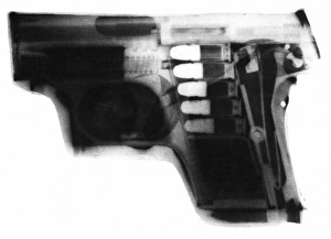
Background imageX Ray Collection: X-RAY OF AUTOMATIC PISTOL. American, 1928

X-RAY OF AUTOMATIC PISTOL. American, 1928

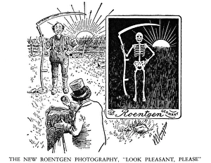
Background imageX Ray Collection: ROENTGEN: X-RAY CARTOON. An American cartoon, c1900, on the discovery of X-rays

ROENTGEN: X-RAY CARTOON. An American cartoon, c1900, on the discovery of X-rays by Wilhelm Conrad Roentgen (1845-1923)

Background imageX Ray Collection: X-ray view of human foot

X-ray view of human foot

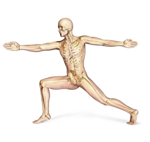
Background imageX Ray Collection: Human male in athletic dynamic posture, with skeleton superimposed

Human male in athletic dynamic posture, with skeleton superimposed
Human male in athletic dynamic posture, with full skeleton superimposed

Background imageX Ray Collection: Conceptual image of pain in human foot

Conceptual image of pain in human foot

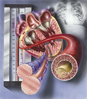
Background imageX Ray Collection: Interior view of heart with detail of muscle cells and atherosclerotic artery

Interior view of heart with detail of muscle cells and atherosclerotic artery. Chest x-ray in background

Background imageX Ray Collection: X-ray image of knee

X-ray image of knee

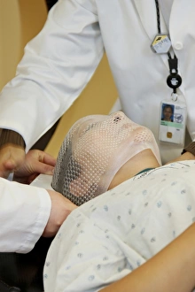
Background imageX Ray Collection: Radiation therapists fit a short face mask to a patient during radiation therapy

Radiation therapists fit a short face mask to a patient during radiation therapy
Two radiation therapists fit a short face mask to a patient model to steady the head during radiation therapy

Background imageX Ray Collection: Female body sitting in dynamic posture with circulatory system superimposed

Female body sitting in dynamic posture with circulatory system superimposed
Human female body sitting in dynamic posture, with full circulatory system superimposed

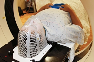
Background imageX Ray Collection: Patient positioned in computed tomography scanner

Patient positioned in computed tomography scanner
A patient model wearing a short face mask is positioned in a computed tomography (CT) scanner to obtain images of a tumor before radiation therapy

Background imageX Ray Collection: Back view of female body with skeletal system superimposed

Back view of female body with skeletal system superimposed

Background imageX Ray Collection: X-ray view of human skeleton showing teeth and gums

X-ray view of human skeleton showing teeth and gums

Background imageX Ray Collection: X-ray view of bones in human hand

X-ray view of bones in human hand

Background imageX Ray Collection: X-ray view of pain in human foot

X-ray view of pain in human foot

Background imageX Ray Collection: Conceptual image of rheumatoid arthritis in the human hand

Conceptual image of rheumatoid arthritis in the human hand
Conceptual image of rheumatoid arthritis (RA) in the human hand. RA is a chronic inflammatory disorder that typically affects the small joints in your hands and feet

Background imageX Ray Collection: W. C. ROENTGEN (1845-1923). German physicist: oil over a photograph

W. C. ROENTGEN (1845-1923). German physicist: oil over a photograph

Background imageX Ray Collection: WILHELM CONRAD ROENTGEN (1845-1923). German physicist. An American cartoon, c1900

WILHELM CONRAD ROENTGEN (1845-1923). German physicist. An American cartoon, c1900, on Roentgens discovery of X-rays

Background imageX Ray Collection: ROENTGEN X-RAY, 1896. The first published X-ray photograph, as reproduced in Wilhelm

ROENTGEN X-RAY, 1896. The first published X-ray photograph, as reproduced in Wilhelm Roentgens paper announcing his accidental discovery of X-rays

Background imageX Ray Collection: X-RAY, 1896. A radiograph, or X-ray photograph, of 1896 revealing shotgun pellets

X-RAY, 1896. A radiograph, or X-ray photograph, of 1896 revealing shotgun pellets in the hand of a New York attorney


All Professionally Made to Order for Quick Shipping

"Unveiling the Hidden World: Exploring the Marvels of X-ray Imaging" Pillars of Creation: Witness the breathtaking beauty of celestial formations through an X-ray lens, revealing a mesmerizing view beyond what meets the eye. Normal Knees, X-rayed: Delve into the intricate structure of our joints as we peer beneath the surface to understand how our knees function in perfect harmony. Panoramic Dental X-ray: Step into a world where dentistry meets technology, capturing a comprehensive image that allows for precise diagnosis and treatment planning. Person with a Camera, X-ray Vision: Unleash your inner superhero as we explore how modern photography techniques merge with X-rays to capture stunning images from within. Tendinitis of the Shoulder, Exposed by X-ray: Discover how this powerful imaging tool helps healthcare professionals identify and diagnose shoulder conditions like tendinitis with accuracy and efficiency. Total Hip Replacement Revealed by X-ray: Journey through medical advancements as we uncover how an artificial hip joint is seamlessly integrated into one's body using cutting-edge surgical techniques guided by detailed x-rays. Normal Skull Underneath It All - An Intriguing Peek Through an X-ray Lens: Explore the hidden secrets concealed within our skulls as we unravel their fascinating anatomy through x-rays. Tuberculosis Unmasked - A Diagnostic Breakthrough via X-rays: Shedding light on one of humanity's oldest diseases, witness how tuberculosis is detected and monitored using specialized x-rays that aid in early detection and effective treatment strategies. Spiral Galaxy M81 - A Cosmic Symphony Captured in Composite Image Using Advanced Radiography Techniques: Embark on an interstellar journey to marvel at this captivating spiral galaxy captured through innovative composite imaging methods involving x-rays and other astronomical data sources. Skeletons Transformed - Artistic Expression Meets Medical Insight in Striking X-ray Artwork.