Fraud Blocker Skip to main content

X Ray Machine Collection (page 8)

The incredible versatility of the x-ray machine never ceases to amaze us

Background imageX Ray Machine Collection: Large bowel obstruction, X-ray C017 / 7855

Large bowel obstruction, X-ray C017 / 7855
Large bowel obstruction. X-ray of the abdomen of a patient with an obstructed large intestine. The obstruction may be due to cancer, twisting of the intestine, an injury or an infection

Background imageX Ray Machine Collection: Large bowel obstruction, X-ray C017 / 7854

Large bowel obstruction, X-ray C017 / 7854
Large bowel obstruction. X-ray of the abdomen of a patient with an obstructed large intestine. The obstruction may be due to cancer, twisting of the intestine, an injury or an infection

Background imageX Ray Machine Collection: Kidney stones, X-ray C017 / 7856

Kidney stones, X-ray C017 / 7856
Kidney stones. X-ray of the kidney of a patient with kidney stones (dark areas) in the renal pelvis (upper centre) of the kidney

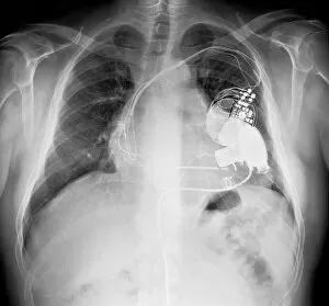
Background imageX Ray Machine Collection: Pacemaker and heart pump, X-ray C017 / 7833

Pacemaker and heart pump, X-ray C017 / 7833
Pacemaker and heart pump. X-ray of the chest of a patient who is waiting for a heart and lung transplant. They have a pacemaker and a left ventricular assist device (LVAD) fitted

Background imageX Ray Machine Collection: Diverticulitis C017 / 7835

Diverticulitis C017 / 7835
Diverticulitis. X-ray of the colon of a patient with diverticulitis. A diverticulum is a small pouch-like structure that protrudes out of weak spots in the intestinal wall

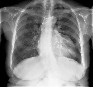
Background imageX Ray Machine Collection: Enlarged heart, X-ray C017 / 7830

Enlarged heart, X-ray C017 / 7830
Enlarged heart. X-ray of the chest of a female patient with an enlarged heart. Cardiac diseases can make the heart need to pump harder

Background imageX Ray Machine Collection: Enlarged heart, X-ray C017 / 7827

Enlarged heart, X-ray C017 / 7827
Enlarged heart. X-ray of the chest of a patient with an enlarged heart and a pacemaker (top right). Cardiac diseases can make the heart need to pump harder

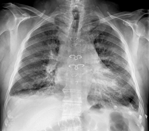
Background imageX Ray Machine Collection: Prosthetic heart valve, X-ray C017 / 7828

Prosthetic heart valve, X-ray C017 / 7828
Prosthetic heart valve. X-ray of the chest of a patient that has had a heart valve replaced with a prosthetic valve. Heart valves control the flow of blood between chambers of the heart

Background imageX Ray Machine Collection: Reversed heart position, X-ray C017 / 7825

Reversed heart position, X-ray C017 / 7825
Reversed heart position. X-ray of the chest of a patient with dextrocardia, the congenital (present at birth) placement of the heart on the right side of the body

Background imageX Ray Machine Collection: Removed lung, X-ray C017 / 7823

Removed lung, X-ray C017 / 7823
Removed lung. X-ray of the chest of a patient that has had their right lung (at left) removed

Background imageX Ray Machine Collection: Lung disease, X-ray C017 / 7817

Lung disease, X-ray C017 / 7817
Lung disease. X-ray of the chest of a patient with chronic obstructive pulmonary disease (COPD). This is a collection of diseases that includes emphysema and chronic bronchitis

Background imageX Ray Machine Collection: Pacemaker, X-ray C017 / 7813

Pacemaker, X-ray C017 / 7813
Pacemaker. X-ray of the chest of a patient that has a pacemaker (upper right) fitted. A pacemaker is a device that supplies electrical impulses to a malfunctioning heart so that it beats normally
Background imageX Ray Machine Collection: Pleural effusion, X-ray C017 / 7811

Pleural effusion, X-ray C017 / 7811
Pleural effusion. X-ray of the chest of a patient with pleural effusion of the left lung (seen on right). Pleural effusion is the accumulation of fluid in the spaces around the lungs
Background imageX Ray Machine Collection: Pleural effusion, X-ray C017 / 7806

Pleural effusion, X-ray C017 / 7806
Pleural effusion. X-ray of the chest of a patient with bilateral pleural effusion. Pleural effusion is the accumulation of fluid in the spaces around the lungs

Background imageX Ray Machine Collection: Dislocated hip replacement, X-ray C017 / 7804

Dislocated hip replacement, X-ray C017 / 7804
Dislocated hip replacement. X-ray of the pelvis of a patient whos total hip replacement of the rigt hip (left) has become dislocated

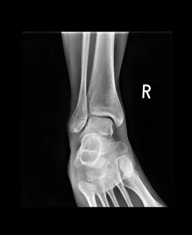
Background imageX Ray Machine Collection: Ankle bone fracture, X-ray C017 / 7809

Ankle bone fracture, X-ray C017 / 7809
Ankle bone fracture. X-ray of the ankle of a patient with a fractured talus (ankle bone)

Background imageX Ray Machine Collection: Dislocated hip replacement, X-ray C017 / 7803

Dislocated hip replacement, X-ray C017 / 7803
Dislocated hip replacement. X-ray of the pelvis of a patient whos total hip replacement of the rigt hip (left) has become dislocated

Background imageX Ray Machine Collection: Ankle bone fracture, X-ray C017 / 7808

Ankle bone fracture, X-ray C017 / 7808
Ankle bone fracture. X-ray of the ankle of a patient with a fractured talus (ankle bone, lower centre)

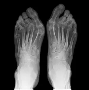
Background imageX Ray Machine Collection: Arthritic feet, X-ray C017 / 7807

Arthritic feet, X-ray C017 / 7807
Arthritic feet. X-ray of the feet of a patient with rheumatoid arthritis. Rheumatoid arthritis is caused by the immune system attacking the bodys own tissues

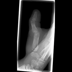
Background imageX Ray Machine Collection: Dislocated thumb, X-ray C017 / 7805

Dislocated thumb, X-ray C017 / 7805
Dislocated thumb. X-ray of the right hand of a 10 year old male patient with a dislocated thumb

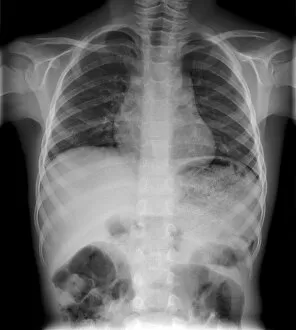
Background imageX Ray Machine Collection: Pulmonary embolism, X-ray C017 / 7802

Pulmonary embolism, X-ray C017 / 7802
Pulmonary embolism. X-ray of the chest of a 72 year old male patient with a pulmonary embolism. This is a blockage of the pulmonary artery

Background imageX Ray Machine Collection: Mesothelioma lung cancer, X-ray C017 / 7776

Mesothelioma lung cancer, X-ray C017 / 7776
Mesothelioma lung cancer, X-ray of the chest of a 84 year old male patient with advanced mesothelioma, a type of lung cancer

Background imageX Ray Machine Collection: Twisted colon, X-ray C017 / 7752

Twisted colon, X-ray C017 / 7752
Twisted colon. X-ray of the abdomen of a 68 year old male patient with a twisted sigmoid colon, a condition known as a volvulus

Background imageX Ray Machine Collection: Pleural effusion, X-ray C017 / 7753

Pleural effusion, X-ray C017 / 7753
Pleural effusion. X-ray of the chest of a 60 year old male patient with pleural effusion of the right lung (seen on left)

Background imageX Ray Machine Collection: Dislocated fingers, X-ray C017 / 7754

Dislocated fingers, X-ray C017 / 7754
Dislocated fingers. X-ray of the hand of a 60 year old male patient who has dislocated his third, fourth and fifth fingers

Background imageX Ray Machine Collection: Swallowed marble, X-ray C017 / 7748

Swallowed marble, X-ray C017 / 7748
Swallowed marble. X-ray of the abdomen of a 4 year old male patient that has swallowed a marble

Background imageX Ray Machine Collection: Swallowed marble, X-ray C017 / 7749

Swallowed marble, X-ray C017 / 7749
Swallowed marble. X-ray of the abdomen of a 4 year old male patient that has swallowed a marble

Background imageX Ray Machine Collection: Broken elbow, X-ray C017 / 7750

Broken elbow, X-ray C017 / 7750
Broken elbow. X-ray of the arm of a 44 year old female patient with a broken elbow

Background imageX Ray Machine Collection: Lung cancer, X-ray C016 / 4435

Lung cancer, X-ray C016 / 4435
Lung cancer. Coloured chest X-ray of a 64 year old male patient with lung cancer. The tumour is seen as the red mass in the right lung (bright area at left)

Background imageX Ray Machine Collection: Lung cancer, X-ray C016 / 4432

Lung cancer, X-ray C016 / 4432
Lung cancer. Coloured chest X-ray of a 64 year old male patient with lung cancer. The tumour is seen as the orange mass in the right lung (dark area at left)

Background imageX Ray Machine Collection: Lung cancer, X-ray C016 / 4433

Lung cancer, X-ray C016 / 4433
Lung cancer. Coloured chest X-ray of a 64 year old male patient with lung cancer. The tumour is seen as the orange mass in the right lung (bright area at left)

Background imageX Ray Machine Collection: Crabs attacking an octopus

Crabs attacking an octopus, coloured X-ray

Background imageX Ray Machine Collection: Crabs and marine molluscs

Crabs and marine molluscs
Crabs and marine mollusc shells, coloured X-ray

Background imageX Ray Machine Collection: X-ray viewing machine, 1895

X-ray viewing machine, 1895
X-ray viewing machine. Historical artwork of the fluoroscope designed and built by the French inventor Eugene Ducretet (1844-1915)

Background imageX Ray Machine Collection: Airport security, explosives detection

Airport security, explosives detection
Airport security officer using a dog to check baggage for explosives. Dogs have an extremely sensitive sense of smell, and can be trained to detect different types of chemicals

Background imageX Ray Machine Collection: Starfish and marine molluscs

Starfish and marine molluscs
Starfish and marine mollusc shells, coloured X- ray

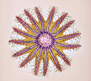
Background imageX Ray Machine Collection: Crown-of-thorns starfish skeleton, X-ray

Crown-of-thorns starfish skeleton, X-ray
Crown-of-thorns starfish skeleton. Coloured X-ray of the crown-of-thorns starfish (Acanthaster planci). This starfish lives on coral reefs in the Indo-Pacific region

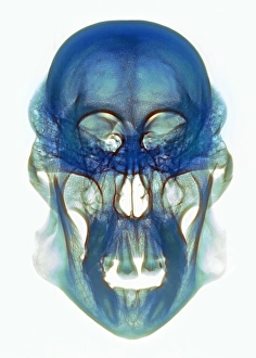
Background imageX Ray Machine Collection: Orangutan skull, X-ray

Orangutan skull, X-ray
Orangutan skull. Coloured X-ray of an orangutan (Pongo sp.) skull. The orangutan is native to Malaysia and Indonesia

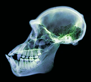
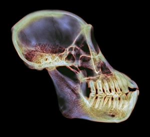
Background imageX Ray Machine Collection: Chimpanzee skull, X-ray

Chimpanzee skull, X-ray
Chimpanzee skull. Coloured X-ray of a side view of a chimpanzees (Pan troglodytes) skull. The cranium (upper right) is considerably smaller than in humans

Background imageX Ray Machine Collection: Orangutan skull

Orangutan skull. Coloured X-ray of an orangutan (Pongo sp.) skull. The orangutan is native to Malaysia and Indonesia

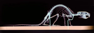
Background imageX Ray Machine Collection: Rat skeleton

Rat skeleton. Coloured X-ray of a rat (Rattus sp.) specimen. The rat is held in place by wire supports (blue vertical lines) which are attached to a base (bottom)

Background imageX Ray Machine Collection: Shark

Shark (class Chondrichthyes). Coloured X-ray of a transverse section through a shark. Its nervous system (yellow) is made up of a brain (upper centre) and spinal column (yellow line)

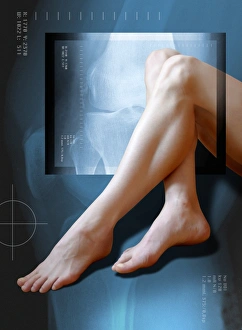
Background imageX Ray Machine Collection: Womans legs, with knee X-ray

Womans legs, with knee X-ray
MODEL RELEASED. Womans legs superimposed on an X-ray of a knee joint. X-rays can be used to diagnose disorders of the skeletal system

Background imageX Ray Machine Collection: Small intestine, barium X-ray

Small intestine, barium X-ray
Small intestines, coloured X-ray. The four X-rays reveal the passage of a swallowed barium contrast medium through the upper part of the digestive system. The medium shows up white on X-rays

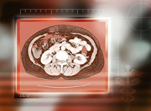
Background imageX Ray Machine Collection: Healthy kidneys, CT scan

Healthy kidneys, CT scan
Healthy kidneys, coloured computed tomography (CT) scan. The kidneys are the two C-shaped white organs either side of the spinal vertebra (centre). Below the spine and kidneys are back muscles

Background imageX Ray Machine Collection: Renal blood vessels, X-ray

Renal blood vessels, X-ray
Renal blood vessels. Angiogram (X-ray) of blood vessels in a healthy kidney. The large artery entering the kidney is the renal artery, which branches into many smaller capillaries inside the kidney

Background imageX Ray Machine Collection: Lung, X-ray

Lung, X-ray
Lung. Coloured bronchography (X-ray) of a healthy human lung. A contrast medium has been added to show the network of airways (green) in the right lung

Background imageX Ray Machine Collection: Oesophagus, X-ray

Oesophagus, X-ray
Oesophagus, coloured X-ray. The oesophagus (orange) is the tube that carries food and drink from the mouth to the stomach. It has been visualised here using a barium swallow


All Professionally Made to Order for Quick Shipping

The incredible versatility of the x-ray machine never ceases to amaze us. From capturing intricate details of brain blood vessels in a 3D angiogram C007 / 1981, to diagnosing a broken wrist bone in X-ray C017 / 7187, this technology has revolutionized medical imaging. But it doesn't stop there. The x-ray machine has even ventured into the animal kingdom, revealing fascinating insights such as a horse's skull or the delicate skeleton of a seahorse. It seems that no creature is too small or too large for this remarkable tool. In addition to its medical applications, the x-ray machine also showcases its playful side. Who would have thought that skeletons could engage in sports? A rugby-playing skeleton and even one enjoying a refreshing drink are just some how this technology can bring humor and creativity to our lives. Not limited to humans and animals, the x-ray machine extends its reach into other fields as well. Panoramic dental X-rays help dentists assess oral health with precision while an individual holding a camera reveals how this invention itself can be captured on film. Furthermore, we witness how the x-ray machine aids in surgical procedures like total hip replacements by providing surgeons with detailed images for precise placement and alignment. Last but not least, let's not forget about exploring what lies beneath our own skin - normal skulls are examined through X-rays allowing us to marvel at the intricacies hidden within each one of us. From healthcare advancements to artistic expressions and scientific discoveries, these glimpses into various aspects of life demonstrate why the x-ray machine remains an invaluable tool that continues to shape our understanding of both human anatomy and beyond.