Fraud Blocker Skip to main content

Virion Collection (page 11)

A virion is a fascinating microscopic entity that plays a crucial role in the world of viruses and infectious diseases

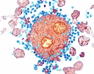
Background imageVirion Collection: Budding HIV particles, TEM C018 / 0125

Budding HIV particles, TEM C018 / 0125
Budding HIV particles. Coloured transmision electron micrograph (TEM) of human immunodeficiency virus (HIV) particles (green) budding from the surface of a T lymphocyte (orange)

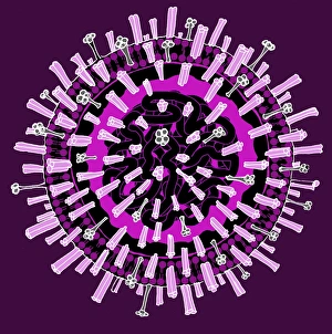
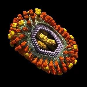
Background imageVirion Collection: Influenza virus, illustration C018 / 0735

Influenza virus, illustration C018 / 0735
Influenza virus. Illustration of an influenza (flu) virus particle (virion). The virus consists of an RNA (ribonucleic acid) core (black)

Background imageVirion Collection: Budding HIV particles, TEM C018 / 0124

Budding HIV particles, TEM C018 / 0124
Budding HIV particles. Coloured transmision electron micrograph (TEM) of human immunodeficiency virus (HIV) particles (gren) budding from the surface of a T lymphocyte (orange) from the H9 cell line

Background imageVirion Collection: Budding HIV particles, TEM C018 / 0123

Budding HIV particles, TEM C018 / 0123
Budding HIV particles. Coloured transmision electron micrograph (TEM) of human immunodeficiency virus (HIV) particles (blue) budding from the surface of a T lymphocyte (red) from the H9 cell line

Background imageVirion Collection: Budding HIV particles, TEM C018 / 0127

Budding HIV particles, TEM C018 / 0127
Budding HIV particles. Coloured transmision electron micrograph (TEM) of human immunodeficiency virus (HIV) particles (orange) budding from the surface of a T lymphocyte (brown)

Background imageVirion Collection: Budding HIV particles, TEM C018 / 0126

Budding HIV particles, TEM C018 / 0126
Budding HIV particles. Coloured transmision electron micrograph (TEM) of human immunodeficiency virus (HIV) particles (pink) budding from the surface of a T lymphocyte (blue) from the H9 cell line

Background imageVirion Collection: SV40 virus capsid, molecular model

SV40 virus capsid, molecular model. Simian virus 40 (SV40) is found in monkeys such as Rhesus monkeys and macaques. Potentially tumour-causing, it is used in laboratory research and in vaccines
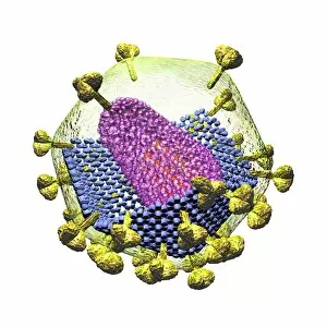
Background imageVirion Collection: Dengue virus capsid, molecular model

Dengue virus capsid, molecular model. This virus, transmitted by mosquito bites, causes the tropical disease dengue fever in humans

Background imageVirion Collection: Ryegrass mottle virus capsid

Ryegrass mottle virus capsid, molecular model. This plant virus is named for its infection of ryegrass, and the discolouration it causes
Background imageVirion Collection: Norwalk virus capsid, molecular model

Norwalk virus capsid, molecular model. This norovirus, which causes a viral form of gastroenteritis, is transmitted from person-to-person or through contaminated food

Background imageVirion Collection: Semliki forest virus capsid

Semliki forest virus capsid, molecular model. This virus, named for the forest in Uganda where it was identified, is spread by the bite of mosquitoes. It can infect both humans and animals

Background imageVirion Collection: Physalis mottle virus capsid

Physalis mottle virus capsid
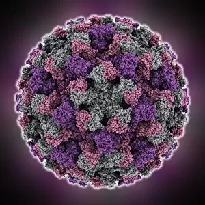
Avian polyomavirus capsid, molecular model. This virus, one of a range named for their potential to cause multiple tumours, infects birds. Discovered in budgerigars in 1981, it is often fatal

Background imageVirion Collection: Bombyx mori densovirus 1 capsid

Bombyx mori densovirus 1 capsid
Bombyx mori densovirus 1 (BmDNV-1), molecular model. This virus infects crustaceans and insects, especially the silkworm (Bombyx mori)

Background imageVirion Collection: Hepatitis B virus capsid, molcular model

Hepatitis B virus capsid, molcular model
Hepatitis B virus capsid, molecular model. This virus, transmitted through infected bodily fluids or blood, causes the disease hepatitis B in humans, leading to acute liver inflammation

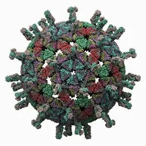
Background imageVirion Collection: Simian rotavirus capsid, molecular model

Simian rotavirus capsid, molecular model. This virus is named for its ability to infect the higher primates (simians). Rotaviruses, transmitted by faecal-oral contact

Background imageVirion Collection: Poliovirus type 3 capsid, molecular model

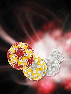
Poliovirus type 3 capsid, molecular model. This enterovirus causes poliomyelitis (polio) in humans, which affects the nervous system, sometimes leading to paralysis

Background imageVirion Collection: Adeno-associated virus capsid

Adeno-associated virus capsid, molecular model. The capsid is a protein shell that encloses the virus genetic information

Background imageVirion Collection: Infectious bursal disease virus capsid

Infectious bursal disease virus capsid, molecular model. This avian virus infects the bursa of Fabricius (specialised bird immune organ) in young chickens, and can cause high mortality rates

Background imageVirion Collection: Enterovirus particle C014 / 4900

Enterovirus particle C014 / 4900
Enterovirus particle. Computer artwork of an enterovirus particle (virion), showing the structure of the capsid (outer shell)

Background imageVirion Collection: Enterovirus capsid proteins structure C014 / 4897

Enterovirus capsid proteins structure C014 / 4897
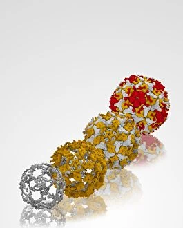
Enterovirus capsid proteins structure. Computer artwork showing how the four component proteins (VP1 to VP4) of an enterovirus particle (virion) interlock to form the capsid (outer shell)

Background imageVirion Collection: Virus, artwork C014 / 4951

Virus, artwork C014 / 4951
Computer artwork of a generic virus particle

Background imageVirion Collection: Enterovirus capsid proteins structure C014 / 4896

Enterovirus capsid proteins structure C014 / 4896
Enterovirus capsid proteins structure. Computer artwork showing how the four component proteins (VP1 to VP4) of an enterovirus particle (virion) interlock to form the capsid (outer shell)

Background imageVirion Collection: Enterovirus particle C014 / 4898

Enterovirus particle C014 / 4898
Enterovirus particle. Computer artwork of an enterovirus particle (virion), showing the structure of the capsid (outer shell)

Background imageVirion Collection: Human poliovirus, molecular model

Human poliovirus, molecular model
Human poliovirus particle. Computer model of the capsid of the human poliovirus. The capsid is a protein coat that encloses the viruss genetic information (genome), stored as RNA (ribonucleic acid)

Background imageVirion Collection: Enterovirus particles C014 / 4899

Enterovirus particles C014 / 4899
Enterovirus particles. Computer artwork of enterovirus particles (virion). Enteroviruses are a genus of non-enveloped positive-sense single-stranded RNA viruses associated with several human

Background imageVirion Collection: HIV particles, artwork C016 / 8659

HIV particles, artwork C016 / 8659
HIV particles, computer artwork. HIV (human immunodeficiency virus) causes AIDS (acquired immune deficiency syndrome). The virus consists of an RNA (ribonucleic acid) genome (pink)

Background imageVirion Collection: HIV particles, artwork C016 / 8658

HIV particles, artwork C016 / 8658
HIV particles, computer artwork. HIV (human immunodeficiency virus) causes AIDS (acquired immune deficiency syndrome). The virus consists of an RNA (ribonucleic acid) genome (pink)

Background imageVirion Collection: HIV particles, artwork C016 / 8655

HIV particles, artwork C016 / 8655
HIV particles, computer artwork. HIV (human immunodeficiency virus) causes AIDS (acquired immune deficiency syndrome). The virus consists of an RNA (ribonucleic acid) genome (pink)

Background imageVirion Collection: HIV particles, artwork C016 / 8654

HIV particles, artwork C016 / 8654
HIV particles, computer artwork. HIV (human immunodeficiency virus) causes AIDS (acquired immune deficiency syndrome). The virus consists of an RNA (ribonucleic acid) genome (pink)

Background imageVirion Collection: HIV particles, artwork C016 / 8652

HIV particles, artwork C016 / 8652
HIV particles, computer artwork. HIV (human immunodeficiency virus) causes AIDS (acquired immune deficiency syndrome). The virus consists of an RNA (ribonucleic acid) genome (pink)

Background imageVirion Collection: HIV particles, artwork C016 / 8653

HIV particles, artwork C016 / 8653
HIV particles, computer artwork. HIV (human immunodeficiency virus) causes AIDS (acquired immune deficiency syndrome). The virus consists of an RNA (ribonucleic acid) genome (pink)

Background imageVirion Collection: HIV particle, artwork C016 / 8649

HIV particle, artwork C016 / 8649
HIV particle, computer artwork. HIV (human immunodeficiency virus) causes AIDS (acquired immune deficiency syndrome). The virus consists of an RNA (ribonucleic acid) genome

Background imageVirion Collection: HIV particle, artwork C016 / 8651

HIV particle, artwork C016 / 8651
HIV particle, computer artwork. HIV (human immunodeficiency virus) causes AIDS (acquired immune deficiency syndrome). The virus consists of an RNA (ribonucleic acid) genome

Background imageVirion Collection: HIV particle, artwork C016 / 8650

HIV particle, artwork C016 / 8650
HIV particle, computer artwork. HIV (human immunodeficiency virus) causes AIDS (acquired immune deficiency syndrome). The virus consists of an RNA (ribonucleic acid) genome

Background imageVirion Collection: HIV particle, artwork C016 / 8648

HIV particle, artwork C016 / 8648
HIV particle, computer artwork. HIV (human immunodeficiency virus) causes AIDS (acquired immune deficiency syndrome). The virus consists of an RNA (ribonucleic acid) genome (pink)

Background imageVirion Collection: HIV particle, artwork C016 / 8647

HIV particle, artwork C016 / 8647
HIV particle, computer artwork. HIV (human immunodeficiency virus) causes AIDS (acquired immune deficiency syndrome). The virus consists of an RNA (ribonucleic acid) genome (pink)

Background imageVirion Collection: HIV particle, artwork C016 / 8646

HIV particle, artwork C016 / 8646
HIV particle, computer artwork. HIV (human immunodeficiency virus) causes AIDS (acquired immune deficiency syndrome). The virus consists of an RNA (ribonucleic acid) genome (pink)

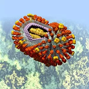
Background imageVirion Collection: Influenza virus, artwork C016 / 8349

Influenza virus, artwork C016 / 8349
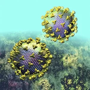
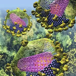
Influenza virus. Cut-away computer artwork of an influenza (flu) virus particle (virion). In each particles lipid envelope (green) are two types of protein spike, haemagglutinin (H)

Background imageVirion Collection: Influenza virus, artwork C016 / 8348

Influenza virus, artwork C016 / 8348
Influenza virus. Cut-away computer artwork of an influenza (flu) virus particle (virion). In each particles lipid envelope (green) are two types of protein spike, haemagglutinin (H)

Background imageVirion Collection: Influenza virus, artwork C016 / 8347

Influenza virus, artwork C016 / 8347
Influenza virus. Cut-away computer artwork of an influenza (flu) virus particle (virion). In each particles lipid envelope (green) are two types of protein spike, haemagglutinin (H)

Background imageVirion Collection: Influenza virus, artwork C016 / 8346

Influenza virus, artwork C016 / 8346
Influenza virus. Cut-away computer artwork of an influenza (flu) virus particle (virion). In each particles lipid envelope (green) are two types of protein spike, haemagglutinin (H)

Background imageVirion Collection: Influenza virus, artwork C016 / 8344

Influenza virus, artwork C016 / 8344
Influenza virus. Cut-away computer artwork of an influenza (flu) virus particle (virion). In each particles lipid envelope (green) are two types of protein spike, haemagglutinin (H)

Background imageVirion Collection: Influenza virus, artwork C016 / 8345

Influenza virus, artwork C016 / 8345
This image may not be used in educational posters Influenza virus. Cut-away computer artwork of an influenza (flu) virus particle (virion)

Background imageVirion Collection: Influenza virus, artwork C016 / 8342

Influenza virus, artwork C016 / 8342
Influenza virus. Computer artwork of an influenza (flu) virus particle (virion). In each particles lipid envelope (green) are two types of protein spike, haemagglutinin (H, red) and neuraminidase (N)

Background imageVirion Collection: Influenza virus, artwork C016 / 8341

Influenza virus, artwork C016 / 8341
Influenza virus. Computer artwork of an influenza (flu) virus particle (virion). In each particles lipid envelope (green) are two types of protein spike, haemagglutinin (H, red) and neuraminidase (N)

Background imageVirion Collection: Influenza virus, artwork C016 / 8340

Influenza virus, artwork C016 / 8340
Influenza virus. Computer artwork of an influenza (flu) virus particle (virion). In each particles lipid envelope (green) are two types of protein spike, haemagglutinin (H, red) and neuraminidase (N)

Background imageVirion Collection: HIV invading white blood cell, artwork

HIV invading white blood cell, artwork
HIV invading white blood cell. Computer artwork showing HIV (human immunodeficiency virus) particles (virions, purple) invading a white blood cell (large, centre) in the human bloodstream

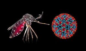
Background imageVirion Collection: Aedes mosquito and Chikungunya virus

Aedes mosquito and Chikungunya virus. Asian tiger mosquito (Aedes albopictus) mosquito next to a Chikungunya virus particle (virion). The Chikungunya virus is transmitted by Aedes sp


All Professionally Made to Order for Quick Shipping

A virion is a fascinating microscopic entity that plays a crucial role in the world of viruses and infectious diseases. From the avian flu virus to HIV particles, norovirus particles, and hepatitis B viruses, these tiny structures are responsible for causing widespread infections. Under the powerful lens of a transmission electron microscope (TEM), we can observe the intricate details of various virions. The avian flu virus appears as an ominous presence with its spiky outer surface, while HIV particles reveal their complex structure that enables them to invade human immune cells. Norovirus particles captured by TEM showcase their round shape and distinctive pattern, reminding us of the havoc they wreak on our digestive system during outbreaks. Sneezing becomes more alarming when we realize it can spread infections like wildfire, as depicted in artwork C013 / 5949. Coronavirus particles also make an appearance under TEM; their crown-like spikes serve as a reminder of how this family of viruses has brought about global health crises such as SARS-CoV-2. Adenoviruses stand out with their geometric shapes portrayed in stunning artwork. Hepatitis B viruses take center stage once again due to their persistence and ability to cause chronic liver disease. Computer-generated artwork showcases influenza virus' unique structure while TEM images capture its spherical form along with other strains. Studying virions provides valuable insights into understanding infectious diseases and developing effective countermeasures against them. These captivating entities remind us of the constant battle between humans and pathogens at a microscopic level.