Fraud Blocker Skip to main content

Vertebral Column Collection

The vertebral column, also known as the backbone or spine, is a crucial structure that provides support and protection to the human body

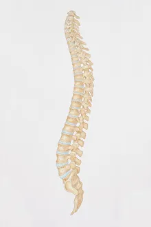
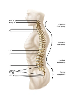
Background imageVertebral Column Collection: Diagram of the human spine, side view

Diagram of the human spine, side view

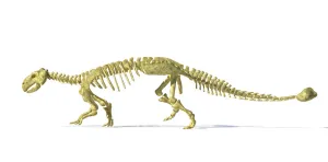
Background imageVertebral Column Collection: 3D rendering of an Ankylosaurus dinosaur skeleton

3D rendering of an Ankylosaurus dinosaur skeleton, side view. This armored dinosaur lived in the early Mesozoic era

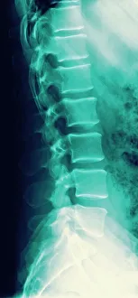
Background imageVertebral Column Collection: Normal spine, X-ray

Normal spine, X-ray
Normal spine. Coloured profile X-ray showing bones of the spine (vertebrae, centre). The front of the body is at right. The ribs (upper right) and the pelvis (bottom) are also seen

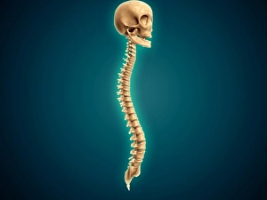
Background imageVertebral Column Collection: Conceptual image of human skull and spinal cord

Conceptual image of human skull and spinal cord

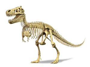
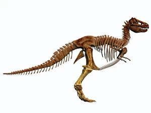
Background imageVertebral Column Collection: 3D rendering of a Tyrannosaurus Rex dinosaur skeleton

3D rendering of a Tyrannosaurus Rex dinosaur skeleton. T-Rex was one of the largest carnivorous dinosaurs of the Cretaceous period

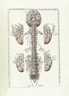
Background imageVertebral Column Collection: The science of human anatomy by Bartholomeo Eustachi

The science of human anatomy by Bartholomeo Eustachi, depicting the shape, size and relative position of the organs of the human body. Circa 18th century

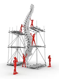
Background imageVertebral Column Collection: Spine with workers, spine repair F007 / 9907

Spine with workers, spine repair F007 / 9907
Conceptual computer artwork depicting a spine being repaired

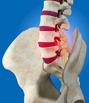
Background imageVertebral Column Collection: Slipped intervertebral disc, artwork

Slipped intervertebral disc, artwork
Slipped intervertebral disc, computer artwork. Intervertebral discs (red) are pads of cartilage between the bones (vertebrae) of the spine

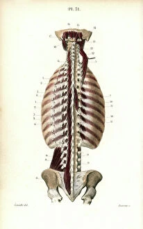
Background imageVertebral Column Collection: Internal muscles of the back, the deepest. Lithograph by Davesne, based on a drawing by Leveille

Internal muscles of the back, the deepest. Lithograph by Davesne, based on a drawing by Leveille
FLO4672659 Internal muscles of the back, the deepest. Lithograph by Davesne, based on a drawing by Leveille, in Petity Atlas complet d'Anatomie descriptive du Corps Humain, by Dr

Background imageVertebral Column Collection: Sympathic nervous system in the thoracic, lumbar and sacral plexus. Lithograph by Davesne

Sympathic nervous system in the thoracic, lumbar and sacral plexus. Lithograph by Davesne
FLO4672529 Sympathic nervous system in the thoracic, lumbar and sacral plexus. Lithograph by Davesne, based on a drawing by Leveille

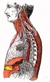
Background imageVertebral Column Collection: Nervous system of the back. Lithograph of Corbie, based on a drawing by Leveille

Nervous system of the back. Lithograph of Corbie, based on a drawing by Leveille
FLO4672457 Nervous system of the back. Lithograph of Corbie, based on a drawing by Leveille, in Petity Atlas complet d'Anatomie descriptive du Corps Humain, by Dr

Background imageVertebral Column Collection: Fern fossil (Pterophyllum sageri). Dinosaur fossil, tooth and vertebra of Nothosaurus

Fern fossil (Pterophyllum sageri). Dinosaur fossil, tooth and vertebra of Nothosaurus, Dracorex tooth
FLO4693297 Fern fossil (Pterophyllum sageri). Dinosaur fossil, tooth and vertebra of Nothosaurus, Dracorex tooth. Fossil fish scales, Gyrolepis, Psamodus, Hybodus, Saurichtys and Acrodus

Background imageVertebral Column Collection: France: Medical photograph of a spinal malformation, 1890

France: Medical photograph of a spinal malformation, 1890
LUX4755402 France: Medical photograph of a spinal malformation, 1890; (add.info.: France: Medical photograph of a spinal malformation, 1890); Photo © Lux-in-Fine.

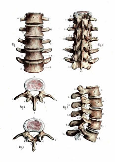
Background imageVertebral Column Collection: Lumbar spine anatomy - 1866 anatomical illustration of the lumbar spine (lower back)

Lumbar spine anatomy - 1866 anatomical illustration of the lumbar spine (lower back) - Lumbar vertebrae
JAB4119471 Lumbar spine anatomy - 1866 anatomical illustration of the lumbar spine (lower back) - Lumbar vertebrae, vertebral column - Osteology, locomotion

Background imageVertebral Column Collection: Thoracic spine anatomy - 1866 anatomical illustration of the thoracic spine

Thoracic spine anatomy - 1866 anatomical illustration of the thoracic spine
JAB4119368 Thoracic spine anatomy - 1866 anatomical illustration of the thoracic spine, showing the individual vertebrae - Vertebral column - Dorsal vertebrae, osteology, locomotion

Background imageVertebral Column Collection: Vertebral column, ribs, vertebrae and sternum. Lithograph of Corbie

Vertebral column, ribs, vertebrae and sternum. Lithograph of Corbie, in Petity Atlas complet d'Anatomie descriptive
FLO4673029 Vertebral column, ribs, vertebrae and sternum. Lithograph of Corbie, in Petity Atlas complet d'Anatomie descriptive du Corps Humain, by Dr

Background imageVertebral Column Collection: Axis and vertebrae. Lithograph of Corbie, in Petity Atlas complet d'Anatomie descriptive du Corps

Axis and vertebrae. Lithograph of Corbie, in Petity Atlas complet d'Anatomie descriptive du Corps Humain, by Dr
FLO4672967 Axis and vertebrae. Lithograph of Corbie, in Petity Atlas complet d'Anatomie descriptive du Corps Humain, by Dr

Background imageVertebral Column Collection: Vertebral column, spine and neck. Lithograph of Corbie, in Petity Atlas complet d'Anatomie

Vertebral column, spine and neck. Lithograph of Corbie, in Petity Atlas complet d'Anatomie descriptive du Corps
FLO4672546 Vertebral column, spine and neck. Lithograph of Corbie, in Petity Atlas complet d'Anatomie descriptive du Corps Humain, by Dr

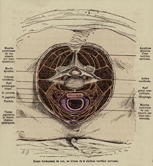
Background imageVertebral Column Collection: Cross-section of the neck at the leval of the sixth cervical vertebra (coloured engraving)

Cross-section of the neck at the leval of the sixth cervical vertebra (coloured engraving)
8655734 Cross-section of the neck at the leval of the sixth cervical vertebra (coloured engraving) by French School, (19th century) (after); Private Collection; (add.info)

Background imageVertebral Column Collection: Sacrum, coccyx and pelvic bone. Lithograph of Corbie, in Petity Atlas complet d'Anatomie

Sacrum, coccyx and pelvic bone. Lithograph of Corbie, in Petity Atlas complet d'Anatomie descriptive du Corps
FLO4673119 Sacrum, coccyx and pelvic bone. Lithograph of Corbie, in Petity Atlas complet d'Anatomie descriptive du Corps Humaine, by Dr

Background imageVertebral Column Collection: Circulatory system of the vertebral column and uterus. Lithograph of Corbie

Circulatory system of the vertebral column and uterus. Lithograph of Corbie, based on a drawing by Leveille
FLO4672977 Circulatory system of the vertebral column and uterus. Lithograph of Corbie, based on a drawing by Leveille, in Petiure Atlas complet d'Anatomie descriptive du Corps Humain, by Dr

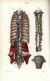
Background imageVertebral Column Collection: Veins of the vertebral column. Lithograph of Corbie, based on a drawing by Leveille

Veins of the vertebral column. Lithograph of Corbie, based on a drawing by Leveille
FLO4672992 Veins of the vertebral column. Lithograph of Corbie, based on a drawing by Leveille, in Petiure Atlas complet d'Anatomie descriptive du Corps Humain, by Dr

Background imageVertebral Column Collection: The circulatory system of the brain has the vertebral column

The circulatory system of the brain has the vertebral column. Lithograph of Anneshower, based on a drawing by Leveille
FLO4672906 The circulatory system of the brain has the vertebral column. Lithograph of Anneshower, based on a drawing by Leveille, in Petiure Atlas complet d'Anatomie descriptive du Corps Humain

Background imageVertebral Column Collection: Spinal bone, ribs and sternum. Lithograph of Corbie, in Petity Atlas complet d'Anatomie

Spinal bone, ribs and sternum. Lithograph of Corbie, in Petity Atlas complet d'Anatomie descriptive du Corps
FLO4672534 Spinal bone, ribs and sternum. Lithograph of Corbie, in Petity Atlas complet d'Anatomie descriptive du Corps Humain, by Dr

Background imageVertebral Column Collection: The thoracic canal. Lithograph of Corbie, based on a drawing by Leveille

The thoracic canal. Lithograph of Corbie, based on a drawing by Leveille, in Petity Atlas complet d'Anatomie
FLO4673048 The thoracic canal. Lithograph of Corbie, based on a drawing by Leveille, in Petity Atlas complet d'Anatomie descriptive du Corps Humain, by Dr

Background imageVertebral Column Collection: Bone of the jaw and neck. Lithograph of Corbie, in Petity Atlas complet d'Anatomie descriptive du

Bone of the jaw and neck. Lithograph of Corbie, in Petity Atlas complet d'Anatomie descriptive du Corps Humain
FLO4672518 Bone of the jaw and neck. Lithograph of Corbie, in Petity Atlas complet d'Anatomie descriptive du Corps Humain, by Dr

Background imageVertebral Column Collection: Aleration and deterioration of internal organs in old age (coloured engraving)

Aleration and deterioration of internal organs in old age (coloured engraving)
8655682 Aleration and deterioration of internal organs in old age (coloured engraving) by French School, (19th century) (after); Private Collection; (add.info)

Background imageVertebral Column Collection: Larynx, trachea and bronchi (coloured engraving)

Larynx, trachea and bronchi (coloured engraving)
8655759 Larynx, trachea and bronchi (coloured engraving) by French School, (19th century) (after); Private Collection; (add.info.: Larynx, trachea and bronchi)

Background imageVertebral Column Collection: Nerves of the tongue and mouth and mechanism of swallowing (coloured engraving)

Nerves of the tongue and mouth and mechanism of swallowing (coloured engraving)
8655754 Nerves of the tongue and mouth and mechanism of swallowing (coloured engraving) by French School, (19th century) (after); Private Collection; (add.info)

Background imageVertebral Column Collection: Cross-section of the head and neck (coloured engraving)

Cross-section of the head and neck (coloured engraving)
8655730 Cross-section of the head and neck (coloured engraving) by French School, (19th century) (after); Private Collection; (add.info.: Cross-section of the head and neck)

Background imageVertebral Column Collection: Great sympathetic nerve - Normal life & health-Drawing A. Demarle 1881

Great sympathetic nerve - Normal life & health-Drawing A. Demarle 1881
JAB4120174 Great sympathetic nerve - Normal life & health-Drawing A. Demarle 1881 by Demarle, Alphonse (1826-1896); (add.info.: Great sympathetic nerve - Normal life & health-Drawing A)

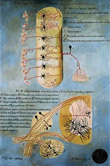
Background imageVertebral Column Collection: Shema histologique des nerfs de la colonne vertebrale. Dessin original en couleurs de Santiago

Shema histologique des nerfs de la colonne vertebrale. Dessin original en couleurs de Santiago Ramon y Cajal (1852-1934)
AIS5385609 Shema histologique des nerfs de la colonne vertebrale. Dessin original en couleurs de Santiago Ramon y Cajal (1852-1934), medecin histologiste espagnol, prix Nobel de medecine en 1906

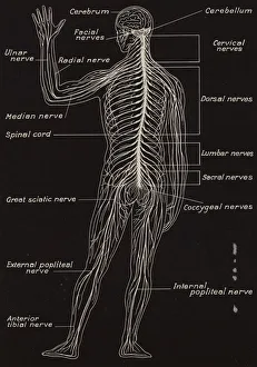
Background imageVertebral Column Collection: Spinal Cord and Spinal Nerves (litho)

Spinal Cord and Spinal Nerves (litho)
7180479 Spinal Cord and Spinal Nerves (litho) by English School, (20th century); Private Collection; (add.info.: Spinal Cord and Spinal Nerves)

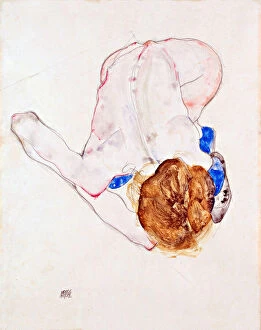
Background imageVertebral Column Collection: Femme nue aux bas bleus, penchee vers l avant - Gouache sur papier de Egon Schiele (1890-1918)

Femme nue aux bas bleus, penchee vers l avant - Gouache sur papier de Egon Schiele (1890-1918)
FIA5386362 Femme nue aux bas bleus, penchee vers l avant - Gouache sur papier de Egon Schiele (1890-1918), 1912 - Nude with Blue Stockings

Background imageVertebral Column Collection: Medicine: reduction of a dislocated spine - patient is fixed on a wooden board

Medicine: reduction of a dislocated spine - patient is fixed on a wooden board
LRI4672796 Medicine: reduction of a dislocated spine - patient is fixed on a wooden board, one of the therapists stands on his back (manuscript, 10th-11th century) by Unknown Artist

Background imageVertebral Column Collection: Anatomical Chart, 1938 (pen and watercolour on cloth laid on canvas)

Anatomical Chart, 1938 (pen and watercolour on cloth laid on canvas)
CHT163244 Anatomical Chart, 1938 (pen and watercolour on cloth laid on canvas) by Tibetan School; Wellcome Museum, London

Background imageVertebral Column Collection: Medicine: reduction of a dislocated spine, patient is hanged with straps to a ladder

Medicine: reduction of a dislocated spine, patient is hanged with straps to a ladder
LRI4672778 Medicine: reduction of a dislocated spine, patient is hanged with straps to a ladder (manuscript, 10th-11th century) by Unknown Artist, (11th century); Biblioteca Medicea-Laurenziana

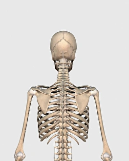
Background imageVertebral Column Collection: Rear view of human skeletal system showing upper back

Rear view of human skeletal system showing upper back

Background imageVertebral Column Collection: Anatomy of human vertebral column, left lateral view

Anatomy of human vertebral column, left lateral view

Background imageVertebral Column Collection: Tyrannosaurus Rex dinosaur skeleton

Tyrannosaurus Rex dinosaur skeleton. T-Rex was an intimidating predator that lived in North America during the Cretaceous Period

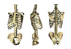
Background imageVertebral Column Collection: Human skeleton anatomy, artwork

Human skeleton anatomy, artwork
Human skeleton anatomy, computer artwork. Three views of the bones of the human torso, seen from an oblique frontal view (left), from the side (centre) and from the front (right)

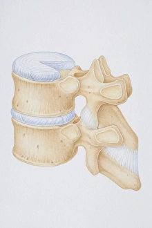
Background imageVertebral Column Collection: Diagram of a human thoracic vertebrae including the intervertebral disk

Diagram of a human thoracic vertebrae including the intervertebral disk, vertebral process and ligaments

Background imageVertebral Column Collection: 3D rendering of human vertebral column with skull

3D rendering of human vertebral column with skull

Background imageVertebral Column Collection: Human spine, side view

Human spine, side view

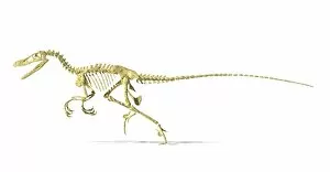
Background imageVertebral Column Collection: 3D rendering of a Velociraptor dinosaur skeleton

3D rendering of a Velociraptor dinosaur skeleton, side view

Background imageVertebral Column Collection: 3D rendering of a Diplodocus dinosaur skeleton

3D rendering of a Diplodocus dinosaur skeleton, side view. Diplodocus was a giant herbivorous dinosaur of the late Jurassic period

Background imageVertebral Column Collection: Spinosaurus dinosaur skeleton

Spinosaurus dinosaur skeleton on textured background

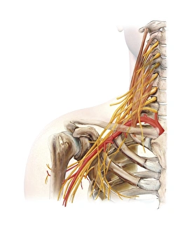
Background imageVertebral Column Collection: Right shoulder and nerve plexus, artwork C016 / 6811

Right shoulder and nerve plexus, artwork C016 / 6811
Right shoulder and nerve plexus. Artwork of the nerves (yellow), arteries (red), and bones of the right shoulder, seen from the front


All Professionally Made to Order for Quick Shipping

The vertebral column, also known as the backbone or spine, is a crucial structure that provides support and protection to the human body. In a diagram of the human spine from a side view, we can see its intricate arrangement of vertebrae stacked on top of each other like building blocks. This 3D rendering reminds us of the remarkable similarity between our own spinal structure and that of an Ankylosaurus dinosaur skeleton. An X-ray image showcases a normal spine, highlighting its flexibility and alignment. The conceptual image featuring a human skull and spinal cord emphasizes the vital connection between these two essential components. Meanwhile, another 3D rendering displays the imposing skeletal frame of a Tyrannosaurus Rex dinosaur, reminding us that even ancient creatures had their own version of this fundamental anatomical feature. Bartholomeo Eustachi's book "The Science of Human Anatomy" serves as an invaluable resource for understanding the intricacies of the vertebral column. It delves into its composition, function, and importance in maintaining overall bodily health. In contrast to scientific illustrations and historical texts lies an artwork depicting workers repairing a damaged spine - F007 / 9907 - symbolizing medical advancements in treating spinal conditions such as slipped intervertebral discs. Another artistic representation by Santiago Ramon y Cajal portrays colorful histological nerves within the vertebral column; his work earned him recognition with a Nobel Prize in Medicine in 1906. Lithographs showcasing the spinal cord and spinal nerves further emphasize their significance in transmitting signals throughout our body. On a different note, Egon Schiele's painting "Nude with Blue Stockings" captures both vulnerability and grace while bending forward – reminding us how every movement involves coordination from our central axis. Lastly, we witness medicine's progress through time with an image illustrating reduction techniques for dislocated spines where patients are secured onto wooden boards during treatment – highlighting how far we have come in providing care and support for spinal injuries.