Fraud Blocker Skip to main content

Vascular Collection (page 5)

"Exploring the Intricate World Anatomy: From Brain Blood Vessels to Maize Roots" Delve into the fascinating realm anatomy

Background imageVascular Collection: Renal blood supply, anatomical artwork

Renal blood supply, anatomical artwork
Renal blood supply. Anatomical computer artwork of a human kidney (left) and adrenal gland (beige, upper centre), showing their main blood supply
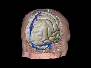
Background imageVascular Collection: Thrombophlebitis in the brain, 3D CT scan

Thrombophlebitis in the brain, 3D CT scan
Thrombophlebitis in the brain. Coloured 3D computed tomography (CT) angiogram of the blood vessels (blue) in the brain of a 48-year-old with thrombophlebitis of the superior sagittal sinus

Background imageVascular Collection: Coronary stenosis after treatment, X-ray

Coronary stenosis after treatment, X-ray
Coronary stenosis after treatment. Angiogram of the left coronary artery in the heart of a 60-year-old patient after balloon catheterisation

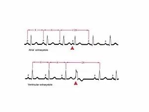
Background imageVascular Collection: Extrasystole heartbeats, ECG artwork

Extrasystole heartbeats, ECG artwork. Diagram showing the electrical activity of the atrium (top) and ventricle (bottom) of a human heart during extrasystole

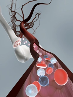
Background imageVascular Collection: Haematopoietic stem cells, artwork

Haematopoietic stem cells, artwork
Haematopoietic stem cells. Cutaway computer artwork showing white blood cells (leucocytes, white, round), red blood cells (erythrocytes, red) and haematopoietic stem cells (HSCs)

Background imageVascular Collection: Ventricular systole, anatomical artwork

Ventricular systole, anatomical artwork
Ventricular systole. Anatomical computer artwork of a transverse section through a healthy human heart during ventricular systole of the cardiac cycle (heartbeat)

Background imageVascular Collection: Stroke, CT scans

Stroke, CT scans
Stroke. Coloured computed tomography (CT) scans of sections through the brain of a patient taken 24 hours, 10 days, 25 days, and 90 days after a haemorrhagic stroke

Background imageVascular Collection: Coronary stenosis treatment, X-rays

Coronary stenosis treatment, X-rays
Coronary stenosis treatment. Coloured angiograms showing the left coronary artery in the heart of a 60-year-old patient before (top left)

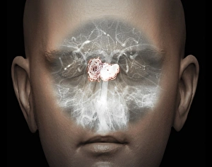
Background imageVascular Collection: Human head, MRI and 3D CT scans

Human head, MRI and 3D CT scans
Stroke. Coloured composite image of a magnetic resonance imaging (MRI) scan of the brain and 2D and 3D computed tomography (CT)

Background imageVascular Collection: Atherosclerosis, 3D CT scan C016 / 6537

Atherosclerosis, 3D CT scan C016 / 6537
Atherosclerosis. Coloured 3D computed tomography (CT) angiogram of the hip of a 67 year old patient with hypertension (high blood pressure) and hypercholesterolemia (high blood cholesterol)

Background imageVascular Collection: Cancer blood supply, conceptual image C016 / 7505

Cancer blood supply, conceptual image C016 / 7505
Cancer blood supply, conceptual image. Computer artwork representing the blood supply to a cancerous (malignant) tumour

Background imageVascular Collection: Atherosclerosis, 3D CT scan C016 / 6540

Atherosclerosis, 3D CT scan C016 / 6540
Atherosclerosis. Coloured 3D computed tomography (CT) angiogram of the abdomen of a 73 year old patient. The aorta (cream, down centre), the bodys main artery, has calcified fatty plaques (atheroma)

Background imageVascular Collection: Atherosclerosis, 3D CT scan C016 / 6499

Atherosclerosis, 3D CT scan C016 / 6499
Atherosclerosis. Coloured 3D computed tomography (CT) angiogram of the head of a 60 year old patient with hypertension (high blood pressure) and hypercholesterolemia (high blood cholesterol)

Background imageVascular Collection: Dicotyledon root, SEM C016 / 9125

Dicotyledon root, SEM C016 / 9125
Dicotyledon root. Coloured scanning electron micrograph (SEM) of a transverse section through an immature root from a dicotyledonous plant

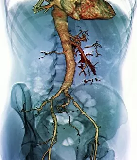
Background imageVascular Collection: Abdominal aorta, 3D CT scan C016 / 6542

Abdominal aorta, 3D CT scan C016 / 6542
Abdominal aorta. Coloured 3D computed tomography (CT) scan of the abdominal aorta of a 56 year old patient. The aorta is the bodys main artery. It carries blood from the heart (at top) around the body

Background imageVascular Collection: Atherosclerosis, 3D CT scan C016 / 6538

Atherosclerosis, 3D CT scan C016 / 6538
Atherosclerosis. Coloured 3D computed tomography (CT) angiogram of the hip of a 67 year old patient with hypertension (high blood pressure) and hypercholesterolemia (high blood cholesterol)

Background imageVascular Collection: Atherosclerosis, 3D CT scan C016 / 6539

Atherosclerosis, 3D CT scan C016 / 6539
Atherosclerosis. Coloured 3D computed tomography (CT) angiogram of the heart of a 69 year old patient that has had an aortic heart valve replacement

Background imageVascular Collection: Dicotyledon root, SEM C016 / 9124

Dicotyledon root, SEM C016 / 9124
Dicotyledon root. Coloured scanning electron micrograph (SEM) of a transverse section through an immature root from a dicotyledonous plant

Background imageVascular Collection: Portal vein surgery, X-ray C016 / 6546

Portal vein surgery, X-ray C016 / 6546
Vein surgery. Coloured angiogram (X-ray) of surgery to lower the blood pressure in the portal vein of a 60 year old patient

Background imageVascular Collection: Placing a catheter, artwork C016 / 6992

Placing a catheter, artwork C016 / 6992
Placing a catheter. Artwork showing the placement of a catheter in a patients blood vessel. A hypodermic needle is being inserted at an angle of 25 degrees (centre left)

Background imageVascular Collection: Dicotyledon root, SEM C016 / 9123

Dicotyledon root, SEM C016 / 9123
Dicotyledon root. Coloured scanning electron micrograph (SEM) of a transverse section through an immature root from a dicotyledonous plant

Background imageVascular Collection: Portal vein surgery, X-ray C016 / 6543

Portal vein surgery, X-ray C016 / 6543
Vein surgery. Angiogram (X-ray) of surgery to lower the blood pressure in the portal vein of a 60 year old patient. A catheter (white line running down from top centre)

Background imageVascular Collection: Shark heart-gill anatomy, artwork C016 / 6847

Shark heart-gill anatomy, artwork C016 / 6847
Shark heart-gill anatomy. Artwork of the heart and gill anatomy of a shark, seen from the underside, with the head at right (teeth at centre right)

Background imageVascular Collection: Skin structure, artwork C016 / 7541

Skin structure, artwork C016 / 7541
Skin structure. Computer artwork showing the different layers and structure of human skin. The upper layer (centre) is the epidermis, with the dermis below

Background imageVascular Collection: Portal vein surgery, X-ray C016 / 6545

Portal vein surgery, X-ray C016 / 6545
Vein surgery. Coloured angiogram (X-ray) of surgery to lower the blood pressure in the portal vein of a 60 year old patient

Background imageVascular Collection: Portal vein surgery, X-ray C016 / 6544

Portal vein surgery, X-ray C016 / 6544
Vein surgery. Coloured angiogram (X-ray) of surgery to lower the blood pressure in the portal vein of a 60 year old patient

Background imageVascular Collection: Male skeleton and organs, artwork

Male skeleton and organs, artwork
Male skeleton and organs and blood vessels, artwork

Background imageVascular Collection: Cranium anatomy, artwork

Cranium anatomy, artwork
Cranium anatomy, computer artwork. At bottom is the brain, which is enclosed in three membranes (grey) known as the meninges

Background imageVascular Collection: Glomerulus, TEM

Glomerulus, TEM
Glomerulus. Transmission electron micrograph (TEM) of a section through a nephron in a human kidney, showing a cluster of capillaries within a renal corpuscle, also known as a glomerulus

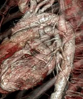
Background imageVascular Collection: Narrowed aorta, 3D CT scans

Narrowed aorta, 3D CT scans
Narrowed aorta. Coloured 3D computed tomography (CT) scans of the heart (centre of each scan) and major blood vessels of a 50-year-old patient

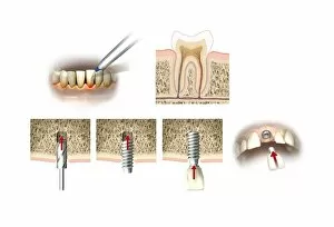
Background imageVascular Collection: Dental implant, artwork

Dental implant, artwork
Dental implant. Artworks showing the process of replacing a tooth (incisor) with a dental implant. At upper left, front lower teeth and a dental instrument are shown in a case of receding

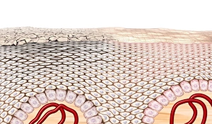
Background imageVascular Collection: Skin anatomy, artwork

Skin anatomy, artwork
Skin anatomy. Artwork of a section through human skin, showing the flattening of skin cells as they rise to the dead keratinised surface layer (top) from the living layer below (the dermis)

Background imageVascular Collection: Calcified heart valve, CT scan

Calcified heart valve, CT scan
Calcified heart valve. Coloured computed tomography (CT) scan of a section through the heart of a 67-year-old patient showing calcification of the aortic valve (round)

Background imageVascular Collection: Stroke, MRI scan

Stroke, MRI scan
Stroke. Coloured magnetic resonance imaging (MRI, left) and computed tomography (CT, right) scans of axial sections through the brain of a 58-year-old patient

Background imageVascular Collection: Berry aneurysm, MRI scan

Berry aneurysm, MRI scan
Stroke. Coloured computed tomography (CT) scan of an axial section through the brain of a 58-year-old patient, showing the damage (dark) caused 24 hours after a cerebral infarction

Background imageVascular Collection: Stroke, CT scan

Stroke, CT scan
Stroke. Coloured computed tomography (CT) scan of an axial section through the brain of a 58-year-old patient, showing the damage (purple) caused 24 hours after a cerebral infarction

Background imageVascular Collection: Calcified heart valve, CT scans

Calcified heart valve, CT scans
Calcified heart valve. Coloured computed tomography (CT) scans of a section through the heart of a 67-year-old patient showing calcification of the aortic valve (round)

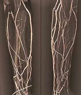
Background imageVascular Collection: Forearm veins, X-ray C018 / 0475

Forearm veins, X-ray C018 / 0475
Forearm veins. Coloured venogram (vein X-ray) of the veins in the forearms of a 43 year old patient in need of dialysis. This venogram has been taken to evaluate the health of the veins prior to

Background imageVascular Collection: Brain haemorrhage, CT scans C018 / 0569

Brain haemorrhage, CT scans C018 / 0569
Brain haemorrhage. Coloured computed tomography (CT) scans of the brain of a 49 year old patient with a subdural haematoma (blue)

Background imageVascular Collection: Aneurysm treatment, angiogram C018 / 0537

Aneurysm treatment, angiogram C018 / 0537
Aneurysm treatment. Coloured angiogram (X-ray) overlaid on a computer model of the treatment of a 39 year old patient with an aneurysm of the posterior communicating artery

Background imageVascular Collection: Aneurysm treatment, angiogram C018 / 0536

Aneurysm treatment, angiogram C018 / 0536
Aneurysm treatment. Angiogram (X-ray) overlaid on a computer model of the treatment of a 39 year old patient with an aneurysm of the posterior communicating artery

Background imageVascular Collection: Aneurysm treatment, angiogram C018 / 0525

Aneurysm treatment, angiogram C018 / 0525
Aneurysm treatment. Coloured angiogram (X-ray) overlaid on a computer model of the treatment of a patient with an aneurysm of the internal carotid artery

Background imageVascular Collection: Retina layers, SEM C018 / 0521

Retina layers, SEM C018 / 0521
Retina layers, coloured scanning electron micrograph (SEM). The retina of the human eye is found on the inside of the eyeball and is formed from a number of layers

Background imageVascular Collection: Aneurysm treatment, angiogram C018 / 0514

Aneurysm treatment, angiogram C018 / 0514
Aneurysm treatment. Coloured angiogram (X-ray) overlaid on a computer model of the treatment of a 39 year old patient with an aneurysm of the posterior communicating artery

Background imageVascular Collection: Aneurysm treatment, angiogram C018 / 0519

Aneurysm treatment, angiogram C018 / 0519
Aneurysm treatment. Angiogram (X-ray) overlaid on a computer model of the treatment of a patient with an aneurysm of the internal carotid artery

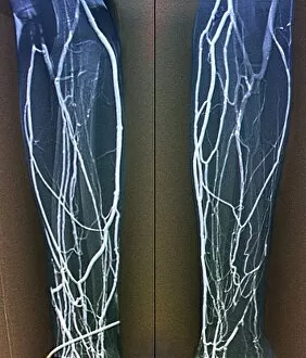
Background imageVascular Collection: Forearm veins, X-ray C018 / 0474

Forearm veins, X-ray C018 / 0474
Forearm veins. Coloured venogram (vein X-ray) of the veins in the forearms of a 43 year old patient in need of dialysis. This venogram has been taken to evaluate the health of the veins prior to

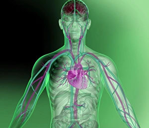
Background imageVascular Collection: Heart and brain vascular system, artwork

Heart and brain vascular system, artwork
Heart and brain vascular system. Artwork showing the arteries (pink) and veins (blue) of the torso, arms, neck, head and brain. The brain is at top (red) and the heart at centre

Background imageVascular Collection: Angiology; systems of veins and arteries C017 / 3462

Angiology; systems of veins and arteries C017 / 3462
Plate from 18th century encyclopedia showing an illustration of the systems of veins and arteries in the field of angiology


All Professionally Made to Order for Quick Shipping

"Exploring the Intricate World Anatomy: From Brain Blood Vessels to Maize Roots" Delve into the fascinating realm anatomy, where a complex network of blood vessels ensures our bodies function seamlessly. Witness the marvels of this intricate system through various historical artworks and scientific imagery. Travel back in time to 1981 with a mesmerizing 3D angiogram C007, showcasing the intricate web of brain blood vessels. Marvel at how these delicate pathways nourish our most vital organ, ensuring its optimal performance. Step into the world of cardiovascular system exploration as you uncover historical artwork depicting its complexity. Admire exquisite neck anatomy illustrations from the 19th century, capturing every detail with artistic precision. Discover the hidden beauty within human heart anatomy through stunning artwork that showcases its awe-inspiring structure. Dive deep into cell structures that make up this remarkable organ, unraveling its secrets one layer at a time. Journey through time as you explore arterial systems depicted in captivating 18th-century artwork. Be captivated by their elegance and understand their crucial role in maintaining overall health and vitality. Immerse yourself in neck vascular anatomy captured by historical artists who sought to document this enigmatic region's intricacies. Let your eyes trace each artery's path as it weaves through muscles and tissues. Witness X-ray images revealing neck and shoulder arteries' hidden mysteries—an opportunity to glimpse beneath our skin's surface and appreciate nature's design flawlessly crafted for optimum functionality. Marvel at Salvia officinalis or Common Sage plant—a botanical wonder known for promoting healthy circulation—its vibrant leaves symbolizing lifeblood coursing through our veins like an eternal river. Transport yourself back to 1825 when male groin arteries were meticulously illustrated—an homage to medical pioneers who unraveled body secrets long before modern technology existed. Uncover artistry behind blood coagulation cascade depicted in breathtaking artwork C016 / 9873.