
Ultrasound Collection
"Unveiling the Unseen: The Power Technology" From rectal exams to steel testing, it has revolutionized various fields with its remarkable capabilities


All Professionally Made to Order for Quick Shipping
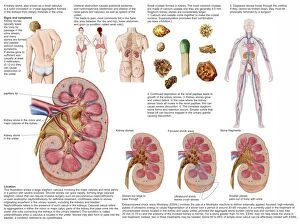
"Unveiling the Unseen: The Power Technology" From rectal exams to steel testing, it has revolutionized various fields with its remarkable capabilities. In 1963, J Beardshaw & Sons in Sheffield, South Yorkshire utilized ultrasonic testing to ensure the quality and integrity of steel. Fast forward to today, doctors in Middlesbrough are harnessing the potential of an advanced ultrasound scanner that can provide unparalleled insights into the human body. One of the most heartwarming applications is seen in prenatal care. A pregnant woman eagerly gazes at her baby scan, capturing a precious moment filled with anticipation and joy. Through cross-sectional biomedical illustrations, we witness how amniocentesis procedures are performed with utmost precision and care. The medical world also benefits from ultrasound's diagnostic prowess when it comes to kidney stones. Comparative images showcase different sizes of these troublesome stones within a human kidney while medical charts meticulously outline their signs and symptoms for accurate diagnosis. Beyond humans, even sheep farming embraces this technology as ewes undergo early-stage pregnancy scans using ultrasound scanners. Farmers gain valuable information about the number of lambs expected, ensuring proper care for both mother and offspring. Obstetric sonography takes center stage during research studies where monitors display intricate details about fetal development. Pregnancy ultrasounds capture tender moments between expectant mothers and their unborn children – treasured memories frozen in time. Ultrasound technology continues to push boundaries by revealing what lies beneath our skin's surface – unlocking mysteries once hidden from view. Its impact spans across industries like medicine, engineering, and agriculture – forever changing how we perceive our world through sound waves transformed into vivid imagery.