
Tunica Externa Collection
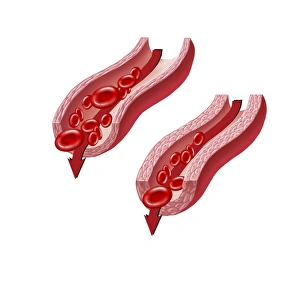
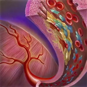
"Tunica Externa: Unveiling the Intricacies of Blood Vessels" Blood flow through a relaxed artery versus an artery in spasm


All Professionally Made to Order for Quick Shipping
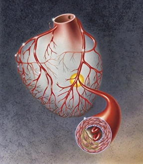
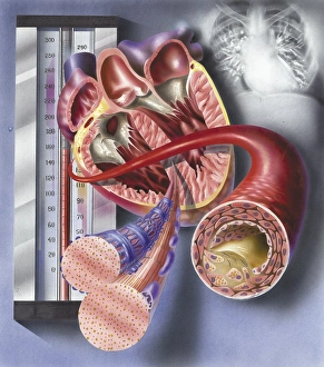
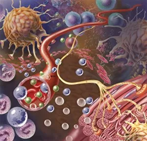
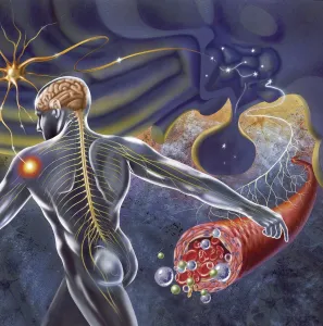
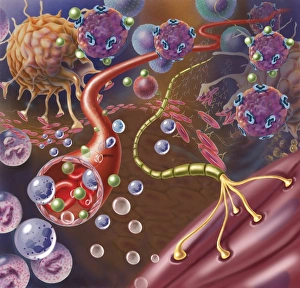
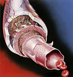
"Tunica Externa: Unveiling the Intricacies of Blood Vessels" Blood flow through a relaxed artery versus an artery in spasm: Witness the contrasting dynamics as blood gracefully courses through a relaxed artery, while an artery in spasm constricts and impedes the vital flow. Arteries on heart showing atherosclerotic plaque in an artery: Delve into the intricate network of arteries within the heart, where we uncover the presence of atherosclerotic plaque, silently threatening cardiovascular health. Interior view of heart with detail of muscle cells and atherosclerotic artery: Peer inside the magnificent organ that is our heart, observing its awe-inspiring muscle cells alongside an unfortunate sight - an atherosclerotic artery battling against compromised blood circulation. Nerve ending, seen in lower right, sends pain message from injured muscle: In this captivating image capturing microscopic details, witness how even at such minuscule levels, nerve endings play their crucial role by transmitting pain signals from injured muscles to our consciousness. Thrombus forming on valve within vein: Astonishingly captured is the formation of a thrombus on one of our body's delicate valves within veins – reminding us that vigilance against clotting disorders is paramount for optimal health. Normal artery compared to plaque and thrombus formation in artery: Comparing side by side reveals stark disparities between normal arteries and those plagued by both arterial plaques and menacing thrombi – emphasizing why preventive measures are essential. Schematic of hypothalamus receiving nerve impulses from the body: Embark upon understanding how our remarkable hypothalamus receives intricate nerve impulses from throughout our body via this illuminating schematic representation. Arteriole with red blood cells, white blood cells, and oxygen: Marvel at this vibrant portrayal showcasing arterioles teeming with life - red blood cells, white blood cells, and oxygen.